2XCR

The 3.5A crystal structure of the catalytic core (B'A' region) of Staphylococcus aureus DNA Gyrase complexed with GSK299423 and DNA


Experimental Data Snapshot

  • Method: X-RAY DIFFRACTION
  • Resolution: 3.50 Å
  • R-Value Free: 0.258 
  • R-Value Work: 0.208 
  • R-Value Observed: 0.210 

wwPDB Validation   3D Report Full Report


Ligand Structure Quality Assessment 


This is version 1.2 of the entry. See complete history


Literature

Type Iia Topoisomerase Inhibition by a New Class of Antibacterial Agents.

Bax, B.D.Chan, P.F.Eggleston, D.S.Fosberry, A.Gentry, D.R.Gorrec, F.Giordano, I.Hann, M.M.Hennessy, A.Hibbs, M.Huang, J.Jones, E.Jones, J.Brown, K.K.Lewis, C.J.May, E.W.Saunders, M.R.Singh, O.Spitzfaden, C.Shen, C.Shillings, A.Theobald, A.F.Wohlkonig, A.Pearson, N.D.Gwynn, M.N.

(2010) Nature 466: 935

  • DOI: https://doi.org/10.1038/nature09197
  • Primary Citation of Related Structures:  
    2XCO, 2XCQ, 2XCR, 2XCS, 2XCT

  • PubMed Abstract: 

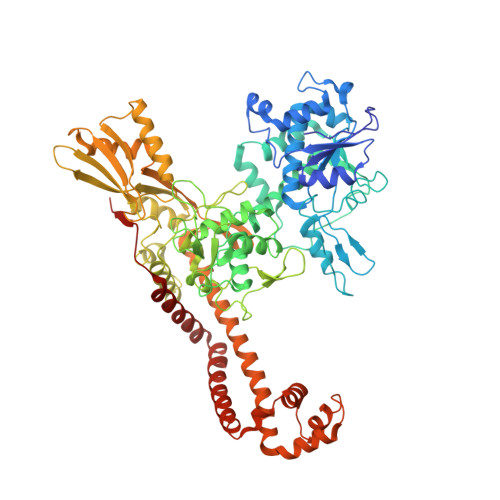
    Despite the success of genomics in identifying new essential bacterial genes, there is a lack of sustainable leads in antibacterial drug discovery to address increasing multidrug resistance. Type IIA topoisomerases cleave and religate DNA to regulate DNA topology and are a major class of antibacterial and anticancer drug targets, yet there is no well developed structural basis for understanding drug action. Here we report the 2.1 A crystal structure of a potent, new class, broad-spectrum antibacterial agent in complex with Staphylococcus aureus DNA gyrase and DNA, showing a new mode of inhibition that circumvents fluoroquinolone resistance in this clinically important drug target. The inhibitor 'bridges' the DNA and a transient non-catalytic pocket on the two-fold axis at the GyrA dimer interface, and is close to the active sites and fluoroquinolone binding sites. In the inhibitor complex the active site seems poised to cleave the DNA, with a single metal ion observed between the TOPRIM (topoisomerase/primase) domain and the scissile phosphate. This work provides new insights into the mechanism of topoisomerase action and a platform for structure-based drug design of a new class of antibacterial agents against a clinically proven, but conformationally flexible, enzyme class.


  • Organizational Affiliation

    Molecular Discovery Research, GlaxoSmithKline, Medicines Research Centre, Gunnels Wood Road, Stevenage, Hertfordshire, SG1 2NY, UK. benjamin.d.bax@gsk.com


Macromolecules

Find similar proteins by:  (by identity cutoff)  |  3D Structure
Entity ID: 1
MoleculeChains Sequence LengthOrganismDetailsImage
DNA GYRASE SUBUNIT B, DNA GYRASE SUBUNIT AA [auth B],
B [auth D],
E [auth S],
F [auth U]
726Staphylococcus aureusMutation(s): 1 
EC: 5.99.1.3
UniProt
Find proteins for Q99XG5 (Staphylococcus aureus (strain N315))
Explore Q99XG5 
Go to UniProtKB:  Q99XG5
Find proteins for P66937 (Staphylococcus aureus (strain N315))
Explore P66937 
Go to UniProtKB:  P66937
Entity Groups  
Sequence Clusters30% Identity50% Identity70% Identity90% Identity95% Identity100% Identity
UniProt GroupsQ99XG5P66937
Sequence Annotations
Expand
  • Reference Sequence

Find similar nucleic acids by:  Sequence   |   3D Structure  

Entity ID: 2
MoleculeChains LengthOrganismImage
5'-D(*5UA*GP*CP*CP*GP*TP*AP*GP*GP*GP*CP*CP*CP*TP*AP*CP*GP *GP*CP*TP)-3'C [auth E],
D [auth F]
20synthetic construct
Sequence Annotations
Expand
  • Reference Sequence

Find similar nucleic acids by:  Sequence   |   3D Structure  

Entity ID: 3
MoleculeChains LengthOrganismImage
5'-D(*AP*GP*CP*CP*GP*TP*AP*GP*GP*GP*CP*CP*CP*TP*AP*CP*GP *GP*CP*TP)-3'G [auth V],
H [auth W]
20synthetic construct
Sequence Annotations
Expand
  • Reference Sequence
Small Molecules
Ligands 1 Unique
IDChains Name / Formula / InChI Key2D Diagram3D Interactions
RXV
Query on RXV

Download Ideal Coordinates CCD File 
I [auth E],
J [auth W]
6-METHOXY-4-(2-{4-[([1,3]OXATHIOLO[5,4-C]PYRIDIN-6-YLMETHYL)AMINO]PIPERIDIN-1-YL}ETHYL)QUINOLINE-3-CARBONITRILE
C25 H27 N5 O2 S
NKXJSCXEAVZSMF-UHFFFAOYSA-N
Experimental Data & Validation

Experimental Data

  • Method: X-RAY DIFFRACTION
  • Resolution: 3.50 Å
  • R-Value Free: 0.258 
  • R-Value Work: 0.208 
  • R-Value Observed: 0.210 
  • Space Group: P 21 21 21
Unit Cell:
Length ( Å )Angle ( ˚ )
a = 113.26α = 90
b = 165.381β = 90
c = 308.431γ = 90
Software Package:
Software NamePurpose
PHENIXrefinement
MOSFLMdata reduction
SCALAdata scaling
PHASERphasing

Structure Validation

View Full Validation Report


Ligand Structure Quality Assessment 


Entry History 

Revision History  (Full details and data files)

  • Version 1.0: 2010-08-04
    Type: Initial release
  • Version 1.1: 2011-05-08
    Changes: Version format compliance
  • Version 1.2: 2011-07-13
    Changes: Version format compliance