2XCK

Crystal structure of PDK1 in complex with a pyrazoloquinazoline inhibitor


Experimental Data Snapshot

  • Method: X-RAY DIFFRACTION
  • Resolution: 2.30 Å
  • R-Value Free: 0.240 
  • R-Value Work: 0.193 
  • R-Value Observed: 0.195 

wwPDB Validation   3D Report Full Report


Ligand Structure Quality Assessment 


This is version 1.3 of the entry. See complete history


Literature

Structure-Based Optimization of Potent Pdk1 Inhibitors.

Angiolini, M.Banfi, P.Casale, E.Casuscelli, F.Fiorelli, C.Saccardo, M.B.Silvagni, M.Zuccotto, F.

(2010) Bioorg Med Chem Lett 20: 4095

  • DOI: https://doi.org/10.1016/j.bmcl.2010.05.070
  • Primary Citation of Related Structures:  
    2XCH, 2XCK

  • PubMed Abstract: 

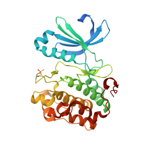
    In this Letter is described the structure-based design of potent dihydro-pyrazoloquinazolines as PDK1 inhibitors. Starting from low potency HTS hits with the aid of X-ray crystallography and modeling, a medicinal chemistry activity was carried out to improve potency versus PDK1 and selectivity versus CDK2 protein kinase.


  • Organizational Affiliation

    Nerviano Medical Sciences Srl, Viale Pasteur 10, 20014 Nerviano, Milano, Italy. mauro.angiolini@nervianoms.com


Macromolecules
Find similar proteins by:  (by identity cutoff)  |  3D Structure
Entity ID: 1
MoleculeChains Sequence LengthOrganismDetailsImage
3-PHOSPHOINOSITIDE DEPENDENT PROTEIN KINASE 1309Homo sapiensMutation(s): 0 
EC: 2.7.11.1
UniProt & NIH Common Fund Data Resources
Find proteins for O15530 (Homo sapiens)
Explore O15530 
Go to UniProtKB:  O15530
PHAROS:  O15530
GTEx:  ENSG00000140992 
Entity Groups  
Sequence Clusters30% Identity50% Identity70% Identity90% Identity95% Identity100% Identity
UniProt GroupO15530
Sequence Annotations
Expand
  • Reference Sequence
Small Molecules
Ligands 3 Unique
IDChains Name / Formula / InChI Key2D Diagram3D Interactions
MH4
Query on MH4

Download Ideal Coordinates CCD File 
B [auth A]1-METHYL-8-{[4-(4-METHYLPIPERAZIN-1-YL)PHENYL]AMINO}-N-[(2-METHYLPYRIDIN-4-YL)METHYL]-4,5-DIHYDRO-1H-PYRAZOLO[4,3-H]QUINAZOLINE-3-CARBOXAMIDE
C29 H33 N9 O
BMPXFYMECYEYOL-UHFFFAOYSA-N
SO4
Query on SO4

Download Ideal Coordinates CCD File 
G [auth A],
H [auth A],
I [auth A]
SULFATE ION
O4 S
QAOWNCQODCNURD-UHFFFAOYSA-L
GOL
Query on GOL

Download Ideal Coordinates CCD File 
C [auth A],
D [auth A],
E [auth A],
F [auth A]
GLYCEROL
C3 H8 O3
PEDCQBHIVMGVHV-UHFFFAOYSA-N
Modified Residues  1 Unique
IDChains TypeFormula2D DiagramParent
SEP
Query on SEP
A
L-PEPTIDE LINKINGC3 H8 N O6 PSER
Binding Affinity Annotations 
IDSourceBinding Affinity
MH4 Binding MOAD:  2XCK IC50: 713 (nM) from 1 assay(s)
PDBBind:  2XCK IC50: 713 (nM) from 1 assay(s)
Experimental Data & Validation

Experimental Data

  • Method: X-RAY DIFFRACTION
  • Resolution: 2.30 Å
  • R-Value Free: 0.240 
  • R-Value Work: 0.193 
  • R-Value Observed: 0.195 
  • Space Group: P 32 2 1
Unit Cell:
Length ( Å )Angle ( ˚ )
a = 123.115α = 90
b = 123.115β = 90
c = 47.24γ = 120
Software Package:
Software NamePurpose
REFMACrefinement
DENZOdata reduction
SCALEPACKdata scaling

Structure Validation

View Full Validation Report


Ligand Structure Quality Assessment 


Entry History 

Deposition Data

Revision History  (Full details and data files)

  • Version 1.0: 2010-07-28
    Type: Initial release
  • Version 1.1: 2011-05-08
    Changes: Version format compliance
  • Version 1.2: 2011-07-13
    Changes: Version format compliance
  • Version 1.3: 2023-12-20
    Changes: Data collection, Database references, Derived calculations, Other, Refinement description