A Novel Signal Transduction Protein P(II) Variant from Synechococcus Elongatus Pcc 7942 Indicates a Two-Step Process for Nagk- P(II) Complex Formation.
Fokina, O., Chellamuthu, V.R., Zeth, K., Forchhammer, K.(2010) J Mol Biol 399: 410
- PubMed: 20399792
- DOI: https://doi.org/10.1016/j.jmb.2010.04.018
- Primary Citation of Related Structures:
2XBP - PubMed Abstract:
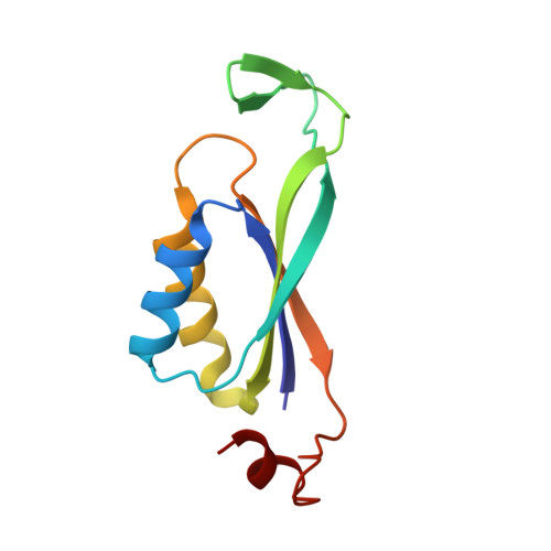
P(II) signal transduction proteins are highly conserved in bacteria, archaea and plants and have key functions in coordination of central metabolism by integrating signals from the carbon, nitrogen and energy status of the cell. In the cyanobacterium Synechococcus elongatus PCC 7942, P(II) binds ATP and 2-oxoglutarate (2-OG) in a synergistic manner, with the ATP binding sites also accepting ADP. Depending on its effector molecule binding status, P(II) (from this cyanobacterium and other oxygenic phototrophs) complexes and regulates the arginine-controlled enzyme of the cyclic ornithine pathway, N-acetyl-l-glutamate kinase (NAGK), to control arginine biosynthesis. To gain deeper insights into the process of P(II) binding to NAGK, we searched for P(II) variants with altered binding characteristics and found P(II) variants I86N and I86T to be able to bind to an NAGK variant (R233A) that was previously shown to be unable to bind wild-type P(II) protein. Analysis of interactions between these P(II) variants and wild-type NAGK as well as with the NAGK R233A variant suggested that the P(II) I86N variant was a superactive NAGK binder. To reveal the structural basis of this property, we solved the crystal structure of the P(II) I86N variant at atomic resolution. The large T-loop, which prevails in most receptor interactions of P(II) proteins, is present in a tightly bended conformation that mimics the T-loop of S. elongatus P(II) after having latched onto NAGK. Moreover, both P(II) I86 variants display a specific defect in 2-OG binding, implying a role of residue I86 in 2-OG binding. We propose a two-step model for the mechanism of P(II)-NAGK complex formation: in an initiating step, a contact between R233 of NAGK and E85 of P(II) initiates the bending of the extended T-loop of P(II), followed by a second step, where a bended T-loop deeply inserts into the NAGK clefts to form the tight complex.
Organizational Affiliation:
Interfakultäres Institut für Mikrobiologie und Infektionsmedizin der Eberhard-Karls-Universität Tübingen, Auf der Morgenstelle 28, 72076 Tübingen, Germany.