2XB8

Structure of Mycobacterium tuberculosis type II dehydroquinase in complex with inhibitor compound (2R)-2-(4-methoxybenzyl)-3- dehydroquinic acid


Experimental Data Snapshot

  • Method: X-RAY DIFFRACTION
  • Resolution: 2.40 Å
  • R-Value Free: 0.206 
  • R-Value Work: 0.156 
  • R-Value Observed: 0.160 

wwPDB Validation   3D Report Full Report


Ligand Structure Quality Assessment 


This is version 1.3 of the entry. See complete history


Literature

Understanding the Key Factors that Control the Inhibition of Type II Dehydroquinase by (2R)-2- Benzyl-3-Dehydroquinic Acids.

Peon, A.Otero, J.M.Tizon, L.Prazeres, V.F.V.Llamas-Saiz, A.L.Fox, G.C.van Raaij, M.J.Lamb, H.Hawkins, A.R.Gago, F.Castedo, L.Gonzalez-Bello, C.

(2010) ChemMedChem 5: 1726

  • DOI: https://doi.org/10.1002/cmdc.201000281
  • Primary Citation of Related Structures:  
    2XB8, 2XB9

  • PubMed Abstract: 

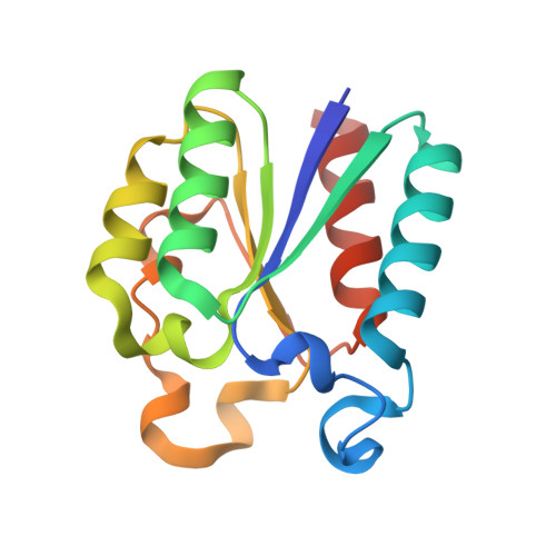
    The binding mode of several substrate analogues, (2R)-2-benzyl-3-dehydroquinic acids 4, which are potent reversible competitive inhibitors of type II dehydroquinase (DHQ2), the third enzyme of the shikimic acid pathway, has been investigated by structural and computational studies. The crystal structures of Mycobacterium tuberculosis and Helicobacter pylori DHQ2 in complex with one of the most potent inhibitor, p-methoxybenzyl derivative 4 a, have been solved at 2.40 Å and 2.75 Å, respectively. This has allowed the resolution of the M. tuberculosis DHQ2 loop containing residues 20-25 for the first time. These structures show the key interactions of the aromatic ring in the active site of both enzymes and additionally reveal an important change in the conformation and flexibility of the loop that closes over substrate binding. The loop conformation and the binding mode of compounds 4 b-d has been also studied by molecular dynamics simulations, which suggest that the benzyl group of inhibitors 4 prevent appropriate orientation of the catalytic tyrosine of the loop for proton abstraction and disrupts its basicity.


  • Organizational Affiliation

    Departamento de Química Orgánica, Universidad de Santiago de Compostela, Santiago de Compostela, Spain.


Macromolecules
Find similar proteins by:  (by identity cutoff)  |  3D Structure
Entity ID: 1
MoleculeChains Sequence LengthOrganismDetailsImage
3-DEHYDROQUINATE DEHYDRATASE146Mycobacterium tuberculosisMutation(s): 0 
EC: 4.2.1.10
UniProt
Find proteins for P9WPX7 (Mycobacterium tuberculosis (strain ATCC 25618 / H37Rv))
Explore P9WPX7 
Go to UniProtKB:  P9WPX7
Entity Groups  
Sequence Clusters30% Identity50% Identity70% Identity90% Identity95% Identity100% Identity
UniProt GroupP9WPX7
Sequence Annotations
Expand
  • Reference Sequence
Small Molecules
Ligands 3 Unique
IDChains Name / Formula / InChI Key2D Diagram3D Interactions
XNW
Query on XNW

Download Ideal Coordinates CCD File 
B [auth A](1R,2R,4S,5R)-1,4,5-TRIHYDROXY-2-(4-METHOXYBENZYL)-3-OXOCYCLOHEXANECARBOXYLIC ACID
C15 H18 O7
KDOXKRDIRCHNDT-YODMDTAWSA-N
TRS
Query on TRS

Download Ideal Coordinates CCD File 
J [auth A]2-AMINO-2-HYDROXYMETHYL-PROPANE-1,3-DIOL
C4 H12 N O3
LENZDBCJOHFCAS-UHFFFAOYSA-O
SO4
Query on SO4

Download Ideal Coordinates CCD File 
C [auth A]
D [auth A]
E [auth A]
F [auth A]
G [auth A]
C [auth A],
D [auth A],
E [auth A],
F [auth A],
G [auth A],
H [auth A],
I [auth A]
SULFATE ION
O4 S
QAOWNCQODCNURD-UHFFFAOYSA-L
Binding Affinity Annotations 
IDSourceBinding Affinity
XNW Binding MOAD:  2XB8 Ki: 26 (nM) from 1 assay(s)
PDBBind:  2XB8 Ki: 26 (nM) from 1 assay(s)
Experimental Data & Validation

Experimental Data

  • Method: X-RAY DIFFRACTION
  • Resolution: 2.40 Å
  • R-Value Free: 0.206 
  • R-Value Work: 0.156 
  • R-Value Observed: 0.160 
  • Space Group: F 2 3
Unit Cell:
Length ( Å )Angle ( ˚ )
a = 126.52α = 90
b = 126.52β = 90
c = 126.52γ = 90
Software Package:
Software NamePurpose
REFMACrefinement
MOSFLMdata reduction
SCALAdata scaling
MOLREPphasing

Structure Validation

View Full Validation Report


Ligand Structure Quality Assessment 


Entry History 

Deposition Data

Revision History  (Full details and data files)

  • Version 1.0: 2010-09-15
    Type: Initial release
  • Version 1.1: 2011-09-28
    Changes: Database references, Refinement description, Version format compliance
  • Version 1.2: 2018-02-07
    Changes: Database references
  • Version 1.3: 2023-12-20
    Changes: Data collection, Database references, Derived calculations, Other, Refinement description