2X9Z

STRUCTURE OF THE PILUS BACKBONE (RRGB) FROM STREPTOCOCCUS PNEUMONIAE


Experimental Data Snapshot

  • Method: X-RAY DIFFRACTION
  • Resolution: 1.30 Å
  • R-Value Free: 0.178 
  • R-Value Work: 0.150 
  • R-Value Observed: 0.152 

wwPDB Validation   3D Report Full Report


This is version 1.3 of the entry. See complete history


Literature

Supramolecular Organization of the Repetitive Backbone Unit of the Streptococcus Pneumoniae Pilus.

Spraggon, G.Koesema, E.Scarselli, M.Malito, E.Biagini, M.Norais, N.Emolo, C.Barocchi, M.A.Giusti, F.Hilleringmann, M.Rappuoli, R.Lesley, S.Covacci, A.Masignani, V.Ferlenghi, I.

(2010) PLoS One 5: 919

  • DOI: https://doi.org/10.1371/journal.pone.0010919
  • Primary Citation of Related Structures:  
    2X9W, 2X9X, 2X9Y, 2X9Z

  • PubMed Abstract: 

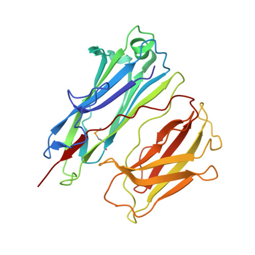
    Streptococcus pneumoniae, like many other Gram-positive bacteria, assembles long filamentous pili on their surface through which they adhere to host cells. Pneumococcal pili are formed by a backbone, consisting of the repetition of the major component RrgB, and two accessory proteins (RrgA and RrgC). Here we reconstruct by transmission electron microscopy and single particle image reconstruction method the three dimensional arrangement of two neighbouring RrgB molecules, which represent the minimal repetitive structural domain of the native pilus. The crystal structure of the D2-D4 domains of RrgB was solved at 1.6 A resolution. Rigid-body fitting of the X-ray coordinates into the electron density map enabled us to define the arrangement of the backbone subunits into the S. pneumoniae native pilus. The quantitative fitting provide evidence that the pneumococcal pilus consists uniquely of RrgB monomers assembled in a head-to-tail organization. The presence of short intra-subunit linker regions connecting neighbouring domains provides the molecular basis for the intrinsic pilus flexibility.


  • Organizational Affiliation

    Genomics Institute of the Novartis Research Foundation, San Diego, California, United States of America. gspraggon@gnf.org


Macromolecules
Find similar proteins by:  (by identity cutoff)  |  3D Structure
Entity ID: 1
MoleculeChains Sequence LengthOrganismDetailsImage
CELL WALL SURFACE ANCHOR FAMILY PROTEIN262Streptococcus pneumoniae TIGR4Mutation(s): 1 
UniProt
Find proteins for A0A0H2UNM7 (Streptococcus pneumoniae serotype 4 (strain ATCC BAA-334 / TIGR4))
Explore A0A0H2UNM7 
Go to UniProtKB:  A0A0H2UNM7
Entity Groups  
Sequence Clusters30% Identity50% Identity70% Identity90% Identity95% Identity100% Identity
UniProt GroupA0A0H2UNM7
Sequence Annotations
Expand
  • Reference Sequence
Small Molecules
Modified Residues  1 Unique
IDChains TypeFormula2D DiagramParent
MSE
Query on MSE
A
L-PEPTIDE LINKINGC5 H11 N O2 SeMET
Experimental Data & Validation

Experimental Data

  • Method: X-RAY DIFFRACTION
  • Resolution: 1.30 Å
  • R-Value Free: 0.178 
  • R-Value Work: 0.150 
  • R-Value Observed: 0.152 
  • Space Group: C 1 2 1
Unit Cell:
Length ( Å )Angle ( ˚ )
a = 94.975α = 90
b = 46.635β = 115.49
c = 64.38γ = 90
Software Package:
Software NamePurpose
PHENIXrefinement
HKL-2000data reduction
SCALEPACKdata scaling
PHASERphasing

Structure Validation

View Full Validation Report


Entry History 

Revision History  (Full details and data files)

  • Version 1.0: 2010-06-30
    Type: Initial release
  • Version 1.1: 2011-05-08
    Changes: Version format compliance
  • Version 1.2: 2011-07-13
    Changes: Version format compliance
  • Version 1.3: 2023-12-20
    Changes: Data collection, Database references, Derived calculations, Other, Refinement description