2X7Y

P450 BM3 F87A in complex with DMSO


Experimental Data Snapshot

  • Method: X-RAY DIFFRACTION
  • Resolution: 2.10 Å
  • R-Value Free: 0.243 
  • R-Value Work: 0.198 
  • R-Value Observed: 0.201 

wwPDB Validation   3D Report Full Report


Ligand Structure Quality Assessment 


This is version 1.4 of the entry. See complete history


Literature

The Role of Active-Site Phe87 in Modulating the Organic Co-Solvent Tolerance of Cytochrome P450 Bm3 Monooxygenase.

Kuper, J.Tee, K.L.Wilmanns, M.Roccatano, D.Schwaneberg, U.Wong, T.S.

(2012) Acta Crystallogr Sect F Struct Biol Cryst Commun 68: 1013

  • DOI: https://doi.org/10.1107/S1744309112031570
  • Primary Citation of Related Structures:  
    2X7Y, 2X80

  • PubMed Abstract: 

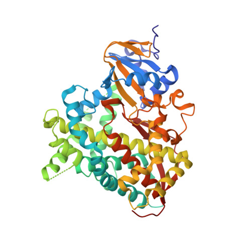
    Understanding the effects of organic co-solvents on protein structure and function is pivotal to engineering enzymes for biotransformation in non-aqueous solvents. The effects of DMSO on the catalytic activity of cytochrome P450 BM3 have previously been investigated and the importance of Phe87 in its organic co-solvent tolerance was identified. To probe the DMSO inactivation mechanism and the functional role of Phe87 in modulating the organic co-solvent tolerance of P450 BM3, the haem domain (Thr1-Leu455) of the F87A variant was cocrystallized in the presence of 14%(v/v) and 28%(v/v) DMSO. At both DMSO concentrations the protein retained the canonical structure of the P450 haem domain without any sign of partial or global unfolding. Interestingly, a DMSO molecule was found in the active site of both structures, with its O atom pointing towards the haem iron. The orientation of the DMSO molecule indicated a dynamic coordination process that was in competition with the active-site water molecule. The ability of the DMSO molecule to coordinate the haem iron is plausibly the main reason why P450 BM3 is inactivated at elevated DMSO concentrations. The data allowed an interesting comparison with the wild-type structures reported previously. A DMSO molecule was found when the wild-type protein was placed in 28%(v/v) DMSO, in which the DMSO molecule coordinated the haem iron directly via its S atom. Intriguingly, no DMSO molecule was observed at 14%(v/v) DMSO for the wild-type structure. These results suggested that the bulky phenyl side chain of Phe87 protects the haem from being accessed by the DMSO molecule and explains the higher tolerance of the wild-type enzyme towards organic co-solvents compared with its F87A variant.


  • Organizational Affiliation

    Rudolf Virchow Centre for Biomedical Research, Josef Schneider Strasse 2, 97070 Würzburg, Germany.


Macromolecules
Find similar proteins by:  (by identity cutoff)  |  3D Structure
Entity ID: 1
MoleculeChains Sequence LengthOrganismDetailsImage
BIFUNCTIONAL P-450/NADPH-P450 REDUCTASE
A, B
455Priestia megateriumMutation(s): 1 
EC: 1.14.14.1
UniProt
Find proteins for P14779 (Priestia megaterium (strain ATCC 14581 / DSM 32 / CCUG 1817 / JCM 2506 / NBRC 15308 / NCIMB 9376 / NCTC 10342 / NRRL B-14308 / VKM B-512 / Ford 19))
Explore P14779 
Go to UniProtKB:  P14779
Entity Groups  
Sequence Clusters30% Identity50% Identity70% Identity90% Identity95% Identity100% Identity
UniProt GroupP14779
Sequence Annotations
Expand
  • Reference Sequence
Small Molecules
Ligands 3 Unique
IDChains Name / Formula / InChI Key2D Diagram3D Interactions
HEM
Query on HEM

Download Ideal Coordinates CCD File 
C [auth A],
H [auth B]
PROTOPORPHYRIN IX CONTAINING FE
C34 H32 Fe N4 O4
KABFMIBPWCXCRK-RGGAHWMASA-L
DMS
Query on DMS

Download Ideal Coordinates CCD File 
G [auth A],
J [auth B]
DIMETHYL SULFOXIDE
C2 H6 O S
IAZDPXIOMUYVGZ-UHFFFAOYSA-N
ZN
Query on ZN

Download Ideal Coordinates CCD File 
D [auth A],
E [auth A],
F [auth A],
I [auth B]
ZINC ION
Zn
PTFCDOFLOPIGGS-UHFFFAOYSA-N
Experimental Data & Validation

Experimental Data

  • Method: X-RAY DIFFRACTION
  • Resolution: 2.10 Å
  • R-Value Free: 0.243 
  • R-Value Work: 0.198 
  • R-Value Observed: 0.201 
  • Space Group: P 21 21 21
Unit Cell:
Length ( Å )Angle ( ˚ )
a = 82.976α = 90
b = 87.88β = 90
c = 159.592γ = 90
Software Package:
Software NamePurpose
REFMACrefinement
MOSFLMdata reduction
SCALAdata scaling
PHASERphasing

Structure Validation

View Full Validation Report


Ligand Structure Quality Assessment 


Entry History 

Deposition Data

Revision History  (Full details and data files)

  • Version 1.0: 2011-03-23
    Type: Initial release
  • Version 1.1: 2012-09-12
    Changes: Database references, Version format compliance
  • Version 1.2: 2012-09-19
    Changes: Database references
  • Version 1.3: 2019-07-24
    Changes: Data collection
  • Version 1.4: 2023-12-20
    Changes: Data collection, Database references, Derived calculations, Other, Refinement description