Structural Insights on the New Mechanism of Trehalose Synthesis by Trehalose Synthase Tret from Pyrococcus Horikoshii.
Woo, E.-J., Ryu, S., Song, H.-N., Jung, T.-Y., Yeon, S., Lee, H., Park, B.C., Park, K., Lee, S.-B.(2010) J Mol Biol 404: 247
- PubMed: 20888836
- DOI: https://doi.org/10.1016/j.jmb.2010.09.056
- Primary Citation of Related Structures:
2X6Q, 2X6R, 2XA1, 2XA2, 2XA9, 2XMP - PubMed Abstract:
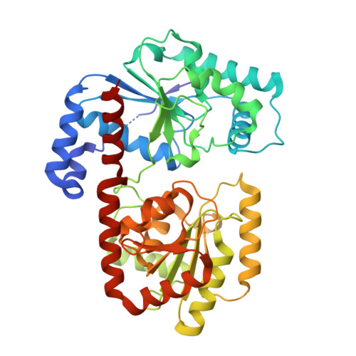
Many microorganisms produce trehalose for stability and survival against various environmental stresses. Unlike the widely distributed trehalose-biosynthetic pathway, which utilizes uridine diphosphate glucose and glucose-6-phosphate, the newly identified enzyme trehalose glycosyltransferring synthase (TreT) from hyperthermophilic bacteria and archaea synthesizes an α,α-trehalose from nucleoside diphosphate glucose and glucose. In the present study, we determined the crystal structure of TreT from Pyrococcus horikoshii at 2.3 Å resolution to understand the detailed mechanism of this novel trehalose synthase. The conservation of essential residues in TreT and the high overall structural similarity of the N-terminal domain to that of trehalose phosphate synthase (TPS) imply that the catalytic reaction of TreT for trehalose synthesis would follow a similar mechanism to that of TPS. The acceptor binding site of TreT shows a wide and commodious groove and lacks the long flexible loop that plays a gating role in ligand binding in TPS. The observation of a wide space at the fissure between two domains and the relative shift of the N-domain in one of the crystal forms suggest that an interactive conformational change between two domains would occur, allowing a more compact architecture for catalysis. The structural analysis and biochemical data in this study provide a molecular basis for understanding the synthetic mechanism of trehalose, or the nucleotide sugar in reverse reaction of the TreT, in extremophiles that may have important industrial implications.
Organizational Affiliation:
Medical Proteomics Research Center, Korea Research Institute of Bioscience and Biotechnology, Daejon 305-806, Republic of Korea. ejwoo@kribb.re.kr