2X5V

80 microsecond laue diffraction snapshot from crystals of a photosynthetic reaction centre 3 millisecond following photoactivation.


Experimental Data Snapshot

  • Method: X-RAY DIFFRACTION
  • Resolution: 3.00 Å
  • R-Value Free: 0.319 
  • R-Value Work: 0.277 
  • R-Value Observed: 0.279 

wwPDB Validation   3D Report Full Report


Ligand Structure Quality Assessment 


This is version 2.1 of the entry. See complete history


Literature

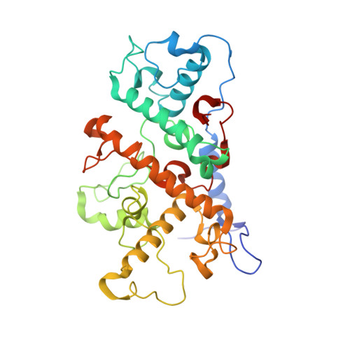
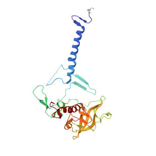
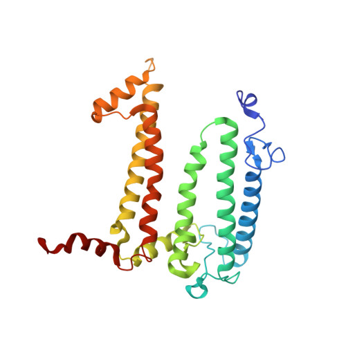
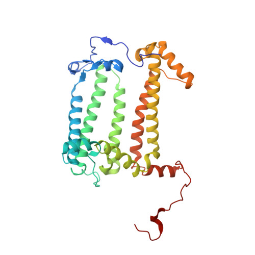
Light-Induced Structural Changes in a Photosynthetic Reaction Center Caught by Laue Diffraction.

Wohri, A.B.Katona, G.Johansson, L.C.Fritz, E.Malmerberg, E.Andersson, M.Vincent, J.Eklund, M.Cammarata, M.Wulff, M.Davidsson, J.Groenhof, G.Neutze, R.

(2010) Science 328: 630

  • DOI: https://doi.org/10.1126/science.1186159
  • Primary Citation of Related Structures:  
    2X5U, 2X5V

  • PubMed Abstract: 

    Photosynthetic reaction centers convert the energy content of light into a transmembrane potential difference and so provide the major pathway for energy input into the biosphere. We applied time-resolved Laue diffraction to study light-induced conformational changes in the photosynthetic reaction center complex of Blastochloris viridis. The side chain of TyrL162, which lies adjacent to the special pair of bacteriochlorophyll molecules that are photooxidized in the primary light conversion event of photosynthesis, was observed to move 1.3 angstroms closer to the special pair after photoactivation. Free energy calculations suggest that this movement results from the deprotonation of this conserved tyrosine residue and provides a mechanism for stabilizing the primary charge separation reactions of photosynthesis.


  • Organizational Affiliation

    Department of Chemical and Biological Engineering, Chalmers University of Technology, Box 462, SE-40530 Göteborg, Sweden.


Macromolecules
Find similar proteins by:  (by identity cutoff)  |  3D Structure
Entity ID: 1
MoleculeChains Sequence LengthOrganismDetailsImage
PHOTOSYNTHETIC REACTION CENTER CYTOCHROME C SUBUNITA [auth C]336Blastochloris viridisMutation(s): 0 
Membrane Entity: Yes 
UniProt
Find proteins for P07173 (Blastochloris viridis)
Explore P07173 
Go to UniProtKB:  P07173
Entity Groups  
Sequence Clusters30% Identity50% Identity70% Identity90% Identity95% Identity100% Identity
UniProt GroupP07173
Sequence Annotations
Expand
  • Reference Sequence
Find similar proteins by:  (by identity cutoff)  |  3D Structure
Entity ID: 2
MoleculeChains Sequence LengthOrganismDetailsImage
REACTION CENTER PROTEIN H CHAINB [auth H]258Blastochloris viridisMutation(s): 0 
Membrane Entity: Yes 
UniProt
Find proteins for P06008 (Blastochloris viridis)
Explore P06008 
Go to UniProtKB:  P06008
Entity Groups  
Sequence Clusters30% Identity50% Identity70% Identity90% Identity95% Identity100% Identity
UniProt GroupP06008
Sequence Annotations
Expand
  • Reference Sequence
Find similar proteins by:  (by identity cutoff)  |  3D Structure
Entity ID: 3
MoleculeChains Sequence LengthOrganismDetailsImage
REACTION CENTER PROTEIN L CHAINC [auth L]274Blastochloris viridisMutation(s): 0 
Membrane Entity: Yes 
UniProt
Find proteins for P06009 (Blastochloris viridis)
Explore P06009 
Go to UniProtKB:  P06009
Entity Groups  
Sequence Clusters30% Identity50% Identity70% Identity90% Identity95% Identity100% Identity
UniProt GroupP06009
Sequence Annotations
Expand
  • Reference Sequence
Find similar proteins by:  (by identity cutoff)  |  3D Structure
Entity ID: 4
MoleculeChains Sequence LengthOrganismDetailsImage
REACTION CENTER PROTEIN M CHAIND [auth M]324Blastochloris viridisMutation(s): 0 
Membrane Entity: Yes 
UniProt
Find proteins for P06010 (Blastochloris viridis)
Explore P06010 
Go to UniProtKB:  P06010
Entity Groups  
Sequence Clusters30% Identity50% Identity70% Identity90% Identity95% Identity100% Identity
UniProt GroupP06010
Sequence Annotations
Expand
  • Reference Sequence
Small Molecules
Ligands 5 Unique
IDChains Name / Formula / InChI Key2D Diagram3D Interactions
BCB
Query on BCB

Download Ideal Coordinates CCD File 
I [auth L],
J [auth L],
L [auth M],
M
BACTERIOCHLOROPHYLL B
C55 H72 Mg N4 O6
QNWPCDKNPGOYNP-DSENBSCCSA-M
BPB
Query on BPB

Download Ideal Coordinates CCD File 
K [auth L],
N [auth M]
BACTERIOPHEOPHYTIN B
C55 H74 N4 O6
SFKCKJXMIAKQMY-GTTFDWDMSA-N
MQ7
Query on MQ7

Download Ideal Coordinates CCD File 
P [auth M]MENAQUINONE-7
C46 H64 O2
RAKQPZMEYJZGPI-LJWNYQGCSA-N
HEC
Query on HEC

Download Ideal Coordinates CCD File 
E [auth C],
F [auth C],
G [auth C],
H [auth C]
HEME C
C34 H34 Fe N4 O4
HXQIYSLZKNYNMH-LJNAALQVSA-N
FE2
Query on FE2

Download Ideal Coordinates CCD File 
O [auth M]FE (II) ION
Fe
CWYNVVGOOAEACU-UHFFFAOYSA-N
Modified Residues  1 Unique
IDChains TypeFormula2D DiagramParent
FME
Query on FME
B [auth H]L-PEPTIDE LINKINGC6 H11 N O3 SMET
Experimental Data & Validation

Experimental Data

  • Method: X-RAY DIFFRACTION
  • Resolution: 3.00 Å
  • R-Value Free: 0.319 
  • R-Value Work: 0.277 
  • R-Value Observed: 0.279 
  • Space Group: P 21 21 2
Unit Cell:
Length ( Å )Angle ( ˚ )
a = 85.693α = 90
b = 143.47β = 90
c = 177.998γ = 90
Software Package:
Software NamePurpose
REFMACrefinement
Precognitiondata reduction
Epinormdata scaling
PHASERphasing

Structure Validation

View Full Validation Report


Ligand Structure Quality Assessment 


Entry History 

Revision History  (Full details and data files)

  • Version 1.0: 2010-05-12
    Type: Initial release
  • Version 1.1: 2012-02-01
    Changes: Non-polymer description, Version format compliance
  • Version 1.2: 2017-01-25
    Changes: Data collection
  • Version 1.3: 2019-05-22
    Changes: Advisory, Data collection, Derived calculations, Experimental preparation
  • Version 2.0: 2019-09-25
    Changes: Atomic model, Data collection, Derived calculations, Non-polymer description, Other, Structure summary
  • Version 2.1: 2023-12-20
    Changes: Advisory, Data collection, Database references, Derived calculations, Refinement description