2X5O

Discovery of Novel 5-Benzylidenerhodanine- and 5-Benzylidene- thiazolidine-2,4-dione Inhibitors of MurD Ligase


Experimental Data Snapshot

  • Method: X-RAY DIFFRACTION
  • Resolution: 1.46 Å
  • R-Value Free: 0.217 
  • R-Value Work: 0.185 
  • R-Value Observed: 0.187 

wwPDB Validation   3D Report Full Report


Ligand Structure Quality Assessment 


This is version 1.3 of the entry. See complete history


Literature

Discovery of novel 5-benzylidenerhodanine and 5-benzylidenethiazolidine-2,4-dione inhibitors of MurD ligase.

Zidar, N.Tomasic, T.Sink, R.Rupnik, V.Kovac, A.Turk, S.Patin, D.Blanot, D.Contreras Martel, C.Dessen, A.Muller Premru, M.Zega, A.Gobec, S.Peterlin Masic, L.Kikelj, D.

(2010) J Med Chem 53: 6584-6594

  • DOI: https://doi.org/10.1021/jm100285g
  • Primary Citation of Related Structures:  
    2WJP, 2X5O

  • PubMed Abstract: 

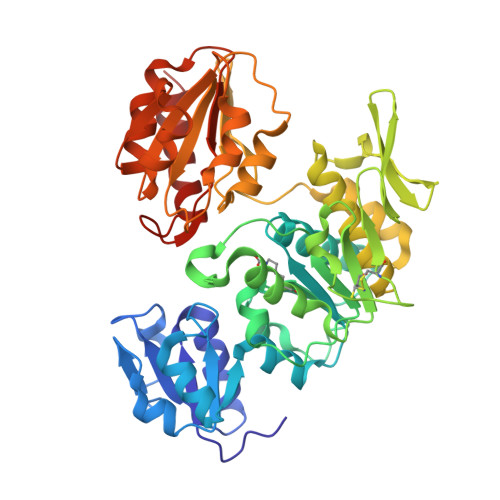
    We have designed, synthesized, and evaluated 5-benzylidenerhodanine- and 5-benzylidenethiazolidine-2,4-dione-based compounds as inhibitors of bacterial enzyme MurD with E. coli IC(50) in the range 45-206 μM. The high-resolution crystal structure of MurD in complex with (R,Z)-2-(3-[{4-([2,4-dioxothiazolidin-5-ylidene]methyl)phenylamino}methyl)benzamido)pentanedioic acid [(R)-32] revealed details of the binding mode of the inhibitor within the active site and provides a good foundation for structure-based design of a novel generation of MurD inhibitors.


  • Organizational Affiliation

    Faculty of Pharmacy, University of Ljubljana, 1000 Ljubljana, Slovenia.


Macromolecules
Find similar proteins by:  (by identity cutoff)  |  3D Structure
Entity ID: 1
MoleculeChains Sequence LengthOrganismDetailsImage
UDP-N-ACETYLMURAMOYLALANINE--D-GLUTAMATE LIGASE439Escherichia coli DH5[alpha]Mutation(s): 0 
EC: 6.3.2.9
UniProt
Find proteins for P14900 (Escherichia coli (strain K12))
Explore P14900 
Go to UniProtKB:  P14900
Entity Groups  
Sequence Clusters30% Identity50% Identity70% Identity90% Identity95% Identity100% Identity
UniProt GroupP14900
Sequence Annotations
Expand
  • Reference Sequence
Small Molecules
Ligands 5 Unique
IDChains Name / Formula / InChI Key2D Diagram3D Interactions
VSV
Query on VSV

Download Ideal Coordinates CCD File 
B [auth A]N-({3-[({4-[(Z)-(2,4-DIOXO-1,3-THIAZOLIDIN-5-YLIDENE)METHYL]PHENYL}AMINO)METHYL]PHENYL}CARBONYL)-D-GLUTAMIC ACID
C23 H21 N3 O7 S
YITBSJALJWUAQA-LVSMMTLPSA-N
SO4
Query on SO4

Download Ideal Coordinates CCD File 
D [auth A],
E [auth A]
SULFATE ION
O4 S
QAOWNCQODCNURD-UHFFFAOYSA-L
SO3
Query on SO3

Download Ideal Coordinates CCD File 
F [auth A]SULFITE ION
O3 S
LSNNMFCWUKXFEE-UHFFFAOYSA-L
AZI
Query on AZI

Download Ideal Coordinates CCD File 
C [auth A]AZIDE ION
N3
IVRMZWNICZWHMI-UHFFFAOYSA-N
CL
Query on CL

Download Ideal Coordinates CCD File 
G [auth A],
H [auth A],
I [auth A],
J [auth A]
CHLORIDE ION
Cl
VEXZGXHMUGYJMC-UHFFFAOYSA-M
Modified Residues  1 Unique
IDChains TypeFormula2D DiagramParent
KCX
Query on KCX
A
L-PEPTIDE LINKINGC7 H14 N2 O4LYS
Binding Affinity Annotations 
IDSourceBinding Affinity
VSV PDBBind:  2X5O IC50: 8.50e+4 (nM) from 1 assay(s)
Binding MOAD:  2X5O IC50: 8.50e+4 (nM) from 1 assay(s)
Experimental Data & Validation

Experimental Data

  • Method: X-RAY DIFFRACTION
  • Resolution: 1.46 Å
  • R-Value Free: 0.217 
  • R-Value Work: 0.185 
  • R-Value Observed: 0.187 
  • Space Group: P 41
Unit Cell:
Length ( Å )Angle ( ˚ )
a = 66.091α = 90
b = 66.091β = 90
c = 135.781γ = 90
Software Package:
Software NamePurpose
REFMACrefinement
XDSdata reduction
XSCALEdata scaling
PHASERphasing

Structure Validation

View Full Validation Report


Ligand Structure Quality Assessment 


Entry History 

Revision History  (Full details and data files)

  • Version 1.0: 2010-09-15
    Type: Initial release
  • Version 1.1: 2011-08-31
    Changes: Database references, Refinement description, Version format compliance
  • Version 1.2: 2018-02-28
    Changes: Database references, Source and taxonomy
  • Version 1.3: 2023-12-20
    Changes: Data collection, Database references, Derived calculations, Other, Refinement description