2X2V

Structural basis of a novel proton-coordination type in an F1Fo-ATP synthase rotor ring


Experimental Data Snapshot

  • Method: X-RAY DIFFRACTION
  • Resolution: 2.50 Å
  • R-Value Free: 0.234 
  • R-Value Work: 0.188 
  • R-Value Observed: 0.190 

wwPDB Validation   3D Report Full Report


Ligand Structure Quality Assessment 


This is version 1.3 of the entry. See complete history


Literature

A New Type of Proton Coordination in an F(1)F(O)- ATP Synthase Rotor Ring.

Preiss, L.Yildiz, O.Hicks, D.B.Krulwich, T.A.Meier, T.

(2010) PLoS Biol 8: 443

  • DOI: https://doi.org/10.1371/journal.pbio.1000443
  • Primary Citation of Related Structures:  
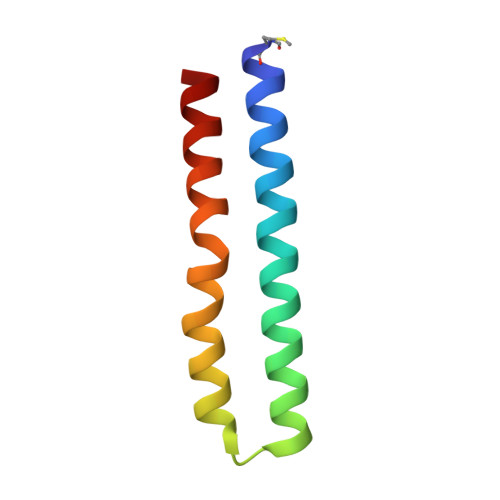
    2X2V

  • PubMed Abstract: 

    We solved the crystal structure of a novel type of c-ring isolated from Bacillus pseudofirmus OF4 at 2.5 A, revealing a cylinder with a tridecameric stoichiometry, a central pore, and an overall shape that is distinct from those reported thus far. Within the groove of two neighboring c-subunits, the conserved glutamate of the outer helix shares the proton with a bound water molecule which itself is coordinated by three other amino acids of outer helices. Although none of the inner helices contributes to ion binding and the glutamate has no other hydrogen bonding partner than the water oxygen, the site remains in a stable, ion-locked conformation that represents the functional state present at the c-ring/membrane interface during rotation. This structure reveals a new, third type of ion coordination in ATP synthases. It appears in the ion binding site of an alkaliphile in which it represents a finely tuned adaptation of the proton affinity during the reaction cycle.


  • Organizational Affiliation

    Department of Structural Biology, Max-Planck Institute of Biophysics, Frankfurt, Germany.


Macromolecules
Find similar proteins by:  (by identity cutoff)  |  3D Structure
Entity ID: 1
MoleculeChains Sequence LengthOrganismDetailsImage
ATP SYNTHASE SUBUNIT C
A, B, C, D, E
A, B, C, D, E, F, G, H, I, J, K, L, M
69Alkalihalophilus pseudofirmus OF4Mutation(s): 0 
Membrane Entity: Yes 
UniProt
Find proteins for P22483 (Alkalihalophilus pseudofirmus (strain ATCC BAA-2126 / JCM 17055 / OF4))
Explore P22483 
Go to UniProtKB:  P22483
Entity Groups  
Sequence Clusters30% Identity50% Identity70% Identity90% Identity95% Identity100% Identity
UniProt GroupP22483
Sequence Annotations
Expand
  • Reference Sequence
Small Molecules
Ligands 2 Unique
IDChains Name / Formula / InChI Key2D Diagram3D Interactions
DPV
Query on DPV

Download Ideal Coordinates CCD File 
AA [auth H]
CA [auth I]
EA [auth J]
GA [auth K]
IA [auth L]
AA [auth H],
CA [auth I],
EA [auth J],
GA [auth K],
IA [auth L],
JA [auth M],
N [auth A],
P [auth B],
R [auth C],
T [auth D],
V [auth E],
X [auth F],
Y [auth G]
dodecyl 2-(trimethylammonio)ethyl phosphate
C17 H38 N O4 P
QBHFVMDLPTZDOI-UHFFFAOYSA-N
NA
Query on NA

Download Ideal Coordinates CCD File 
BA [auth I]
DA [auth J]
FA [auth K]
HA [auth L]
O [auth B]
BA [auth I],
DA [auth J],
FA [auth K],
HA [auth L],
O [auth B],
Q [auth C],
S [auth D],
U [auth E],
W [auth F],
Z [auth H]
SODIUM ION
Na
FKNQFGJONOIPTF-UHFFFAOYSA-N
Modified Residues  1 Unique
IDChains TypeFormula2D DiagramParent
FME
Query on FME
A, B, C, D, E
A, B, C, D, E, F, G, H, I, J, K, L, M
L-PEPTIDE LINKINGC6 H11 N O3 SMET
Experimental Data & Validation

Experimental Data

  • Method: X-RAY DIFFRACTION
  • Resolution: 2.50 Å
  • R-Value Free: 0.234 
  • R-Value Work: 0.188 
  • R-Value Observed: 0.190 
  • Space Group: P 1 21 1
Unit Cell:
Length ( Å )Angle ( ˚ )
a = 74.18α = 90
b = 97.34β = 104.73
c = 121.24γ = 90
Software Package:
Software NamePurpose
PHENIXrefinement
XDSdata reduction
XSCALEdata scaling
PHASERphasing

Structure Validation

View Full Validation Report


Ligand Structure Quality Assessment 


Entry History 

Deposition Data

Revision History  (Full details and data files)

  • Version 1.0: 2010-08-18
    Type: Initial release
  • Version 1.1: 2011-05-07
    Changes: Version format compliance
  • Version 1.2: 2011-07-13
    Changes: Version format compliance
  • Version 1.3: 2023-12-20
    Changes: Data collection, Database references, Derived calculations, Other, Refinement description