2X22

crystal structure of M. tuberculosis InhA inhibited by PT70


Experimental Data Snapshot

  • Method: X-RAY DIFFRACTION
  • Resolution: 2.10 Å
  • R-Value Free: 0.216 
  • R-Value Work: 0.172 
  • R-Value Observed: 0.174 

wwPDB Validation   3D Report Full Report


Ligand Structure Quality Assessment 


This is version 1.3 of the entry. See complete history


Literature

A Slow, Tight Binding Inhibitor of Inha, the Enoyl-Acyl Carrier Protein Reductase from Mycobacterium Tuberculosis.

Luckner, S.R.Liu, N.Am Ende, C.W.Tonge, P.J.Kisker, C.

(2010) J Biol Chem 285: 14330

  • DOI: https://doi.org/10.1074/jbc.M109.090373
  • Primary Citation of Related Structures:  
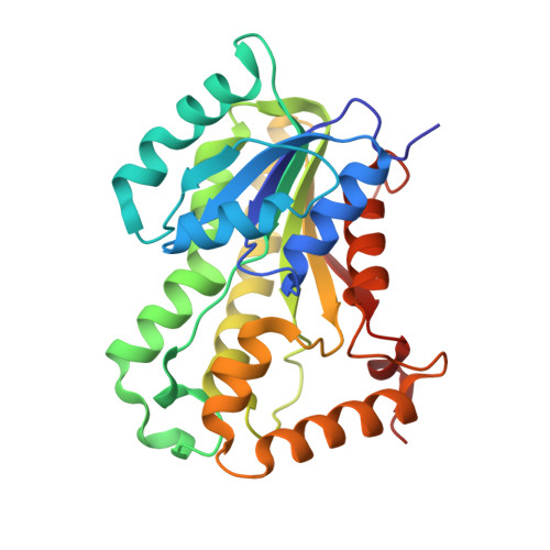
    2X22, 2X23

  • PubMed Abstract: 

    InhA, the enoyl-ACP reductase in Mycobacterium tuberculosis is an attractive target for the development of novel drugs against tuberculosis, a disease that kills more than two million people each year. InhA is the target of the current first line drug isoniazid for the treatment of tuberculosis infections. Compounds that directly target InhA and do not require activation by the mycobacterial catalase-peroxidase KatG are promising candidates for treating infections caused by isoniazid-resistant strains. Previously we reported the synthesis of several diphenyl ethers with nanomolar affinity for InhA. However, these compounds are rapid reversible inhibitors of the enzyme, and based on the knowledge that long drug target residence times are an important factor for in vivo drug activity, we set out to generate a slow onset inhibitor of InhA using structure-based drug design. 2-(o-Tolyloxy)-5-hexylphenol (PT70) is a slow, tight binding inhibitor of InhA with a K(1) value of 22 pm. PT70 binds preferentially to the InhA x NAD(+) complex and has a residence time of 24 min on the target, which is 14,000 times longer than that of the rapid reversible inhibitor from which it is derived. The 1.8 A crystal structure of the ternary complex between InhA, NAD(+), and PT70 reveals the molecular details of enzyme-inhibitor recognition and supports the hypothesis that slow onset inhibition is coupled to ordering of an active site loop, which leads to the closure of the substrate-binding pocket.


  • Organizational Affiliation

    Rudolf Virchow Center for Experimental Biomedicine, Institute for Structural Biology, University of Würzburg, 97080 Würzburg, Germany.


Macromolecules
Find similar proteins by:  (by identity cutoff)  |  3D Structure
Entity ID: 1
MoleculeChains Sequence LengthOrganismDetailsImage
ENOYL-[ACYL-CARRIER-PROTEIN] REDUCTASE [NADH]
A, B
269Mycobacterium tuberculosis H37RvMutation(s): 0 
EC: 1.3.1.9
UniProt
Find proteins for P9WGR1 (Mycobacterium tuberculosis (strain ATCC 25618 / H37Rv))
Explore P9WGR1 
Go to UniProtKB:  P9WGR1
Entity Groups  
Sequence Clusters30% Identity50% Identity70% Identity90% Identity95% Identity100% Identity
UniProt GroupP9WGR1
Sequence Annotations
Expand
  • Reference Sequence
Small Molecules
Binding Affinity Annotations 
IDSourceBinding Affinity
TCU Binding MOAD:  2X22 Ki: 7.8 (nM) from 1 assay(s)
BindingDB:  2X22 Ki: min: 0.02, max: 114 (nM) from 2 assay(s)
IC50: min: 22, max: 590 (nM) from 3 assay(s)
Experimental Data & Validation

Experimental Data

  • Method: X-RAY DIFFRACTION
  • Resolution: 2.10 Å
  • R-Value Free: 0.216 
  • R-Value Work: 0.172 
  • R-Value Observed: 0.174 
  • Space Group: C 2 2 21
Unit Cell:
Length ( Å )Angle ( ˚ )
a = 89.806α = 90
b = 157.513β = 90
c = 91.229γ = 90
Software Package:
Software NamePurpose
REFMACrefinement
MOSFLMdata reduction
TRUNCATEdata scaling
PHASERphasing

Structure Validation

View Full Validation Report


Ligand Structure Quality Assessment 


Entry History 

Deposition Data

Revision History  (Full details and data files)

  • Version 1.0: 2010-03-02
    Type: Initial release
  • Version 1.1: 2011-05-08
    Changes: Version format compliance
  • Version 1.2: 2011-07-13
    Changes: Version format compliance
  • Version 1.3: 2023-12-20
    Changes: Data collection, Database references, Derived calculations, Other, Refinement description