2WZB

The catalytically active fully closed conformation of human phosphoglycerate kinase in complex with ADP, 3PG and magnesium trifluoride


Experimental Data Snapshot

  • Method: X-RAY DIFFRACTION
  • Resolution: 1.47 Å
  • R-Value Free: 0.186 
  • R-Value Work: 0.161 
  • R-Value Observed: 0.162 

wwPDB Validation   3D Report Full Report


Ligand Structure Quality Assessment 


This is version 1.3 of the entry. See complete history


Literature

Transition State Analogue Structures of Human Phosphoglycerate Kinase Establish the Importance of Charge Balance in Catalysis.

Cliff, M.J.Bowler, M.W.Szabo, J.Marston, J.P.M.Varga, A.V.Hownslow, A.M.H.Baxter, N.J.Blackburn, G.M.Vas, M.Waltho, J.P.

(2010) J Am Chem Soc 132: 6507

  • DOI: https://doi.org/10.1021/ja100974t
  • Primary Citation of Related Structures:  
    2WZB, 2WZC, 2WZD

  • PubMed Abstract: 

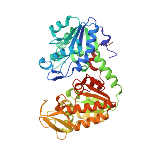
    Transition state analogue (TSA) complexes formed by phosphoglycerate kinase (PGK) have been used to test the hypothesis that balancing of charge within the transition state dominates enzyme-catalyzed phosphoryl transfer. High-resolution structures of trifluoromagnesate (MgF(3)(-)) and tetrafluoroaluminate (AlF(4)(-)) complexes of PGK have been determined using X-ray crystallography and (19)F-based NMR methods, revealing the nature of the catalytically relevant state of this archetypal metabolic kinase. Importantly, the side chain of K219, which coordinates the alpha-phosphate group in previous ground state structures, is sequestered into coordinating the metal fluoride, thereby creating a charge environment complementary to the transferring phosphoryl group. In line with the dominance of charge balance in transition state organization, the substitution K219A induces a corresponding reduction in charge in the bound aluminum fluoride species, which changes to a trifluoroaluminate (AlF(3)(0)) complex. The AlF(3)(0) moiety retains the octahedral geometry observed within AlF(4)(-) TSA complexes, which endorses the proposal that some of the widely reported trigonal AlF(3)(0) complexes of phosphoryl transfer enzymes may have been misassigned and in reality contain MgF(3)(-).


  • Organizational Affiliation

    The Krebs Institute & The Department of Molecular Biology and Biotechnology, The University of Sheffield, Firth Court, Western Bank, Sheffield S10 2TN, UK.


Macromolecules
Find similar proteins by:  (by identity cutoff)  |  3D Structure
Entity ID: 1
MoleculeChains Sequence LengthOrganismDetailsImage
PHOSPHOGLYCERATE KINASE 1416Homo sapiensMutation(s): 0 
EC: 2.7.2.3
UniProt & NIH Common Fund Data Resources
Find proteins for P00558 (Homo sapiens)
Explore P00558 
Go to UniProtKB:  P00558
PHAROS:  P00558
GTEx:  ENSG00000102144 
Entity Groups  
Sequence Clusters30% Identity50% Identity70% Identity90% Identity95% Identity100% Identity
UniProt GroupP00558
Sequence Annotations
Expand
  • Reference Sequence
Small Molecules
Ligands 5 Unique
IDChains Name / Formula / InChI Key2D Diagram3D Interactions
ADP
Query on ADP

Download Ideal Coordinates CCD File 
D [auth A]ADENOSINE-5'-DIPHOSPHATE
C10 H15 N5 O10 P2
XTWYTFMLZFPYCI-KQYNXXCUSA-N
3PG
Query on 3PG

Download Ideal Coordinates CCD File 
E [auth A]3-PHOSPHOGLYCERIC ACID
C3 H7 O7 P
OSJPPGNTCRNQQC-UWTATZPHSA-N
MGF
Query on MGF

Download Ideal Coordinates CCD File 
F [auth A]TRIFLUOROMAGNESATE
F3 Mg
GJOMWUHGUQLOAC-UHFFFAOYSA-K
CL
Query on CL

Download Ideal Coordinates CCD File 
C [auth A]CHLORIDE ION
Cl
VEXZGXHMUGYJMC-UHFFFAOYSA-M
MG
Query on MG

Download Ideal Coordinates CCD File 
B [auth A]MAGNESIUM ION
Mg
JLVVSXFLKOJNIY-UHFFFAOYSA-N
Binding Affinity Annotations 
IDSourceBinding Affinity
3PG BindingDB:  2WZB Kd: 1.09e+5 (nM) from 1 assay(s)
Experimental Data & Validation

Experimental Data

  • Method: X-RAY DIFFRACTION
  • Resolution: 1.47 Å
  • R-Value Free: 0.186 
  • R-Value Work: 0.161 
  • R-Value Observed: 0.162 
  • Space Group: P 21 21 21
Unit Cell:
Length ( Å )Angle ( ˚ )
a = 39.43α = 90
b = 92.18β = 90
c = 109γ = 90
Software Package:
Software NamePurpose
REFMACrefinement
MOSFLMdata reduction
SCALAdata scaling
PHASERphasing

Structure Validation

View Full Validation Report


Ligand Structure Quality Assessment 


Entry History 

Deposition Data

Revision History  (Full details and data files)

  • Version 1.0: 2010-04-14
    Type: Initial release
  • Version 1.1: 2011-07-13
    Changes: Advisory, Version format compliance
  • Version 1.2: 2011-12-07
    Changes: Database references
  • Version 1.3: 2023-12-20
    Changes: Data collection, Database references, Derived calculations, Other, Refinement description