2WYL

Apo structure of a metallo-b-lactamase


Experimental Data Snapshot

  • Method: X-RAY DIFFRACTION
  • Resolution: 2.59 Å
  • R-Value Free: 0.248 
  • R-Value Work: 0.198 
  • R-Value Observed: 0.201 

wwPDB Validation   3D Report Full Report


This is version 1.2 of the entry. See complete history


Literature

Molecular Architecture of the Mn(2+)Dependent Lactonase Ulag Reveals an Rnase-Like Metallo-Beta-Lactamase Fold and a Novel Quaternary Structure.

Garces, F.Fernandez, F.J.Montella, C.Penya-Soler, E.Prohens, R.Aguilar, J.Baldoma, L.Coll, M.Badia, J.Vega, M.C.

(2010) J Mol Biol 398: 715

  • DOI: https://doi.org/10.1016/j.jmb.2010.03.041
  • Primary Citation of Related Structures:  
    2WYL, 2WYM

  • PubMed Abstract: 

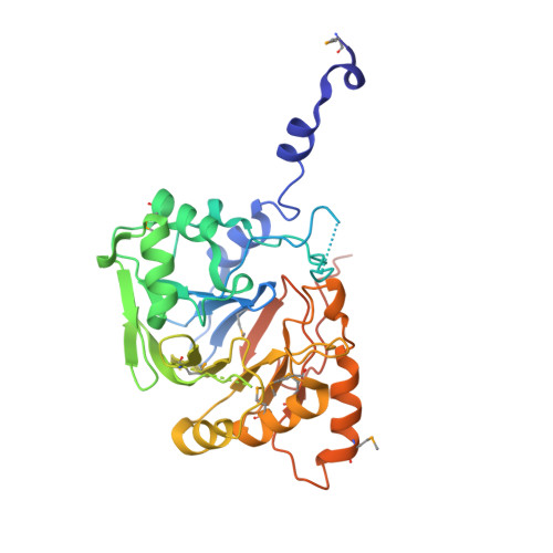
    The ulaG gene, located in the ula regulon, is crucial for the catabolism of l-ascorbate under anaerobic conditions and it has been proposed to encode for the putative l-ascorbate-6-P lactonase. The ulaG gene is widespread among eubacteria, including human commensal and pathogenic genera such as Escherichia, Shigella, Klebsiella and Salmonella. Here, we report the three-dimensional structures of the apoenzyme and Mn(2+) holoenzyme of UlaG from E. coli to 2.6 A resolution, determined using single-wavelength anomalous diffraction phasing and molecular replacement, respectively. The structures reveal a highly specialized metallo-beta-lactamase-like fold derived from an ancient structural template that was involved in RNA maturation and DNA repair. This fold has a novel quaternary architecture consisting of a hexameric ring formed by a trimer of UlaG dimers. A mononuclear Mn(2)(+)-binding site resides at the core of the active site, which displays micromolar affinity for Mn(2+) and a distorted trigonal bipyramidal coordination. The active site Mn(2+) ion can be replaced by Co(2+) or Zn(2+), but not by Fe(3+). We further show that the Mn(2+) or Co(2)(+)-loaded enzyme exhibits lactonase activity towards l-ascorbate 6-P, thereby providing the first direct evidence of its catalytic role in the L-ascorbate catabolic pathway. Guided by the structural homology, we show that UlaG is able to cleave phosphodiester linkages in cyclic nucleotides, suggesting that the conservation of the fold and of the key catalytic residues allows for the evolutionary acquisition of substrate specificity for novel but related substrates.


  • Organizational Affiliation

    Department of Biochemistry and Molecular Biology, Institut de Biomedicina de la Universitat de Barcelona (IBUB), Faculty of Pharmacy, University of Barcelona, Barcelona, ES 08028, Spain.


Macromolecules
Find similar proteins by:  (by identity cutoff)  |  3D Structure
Entity ID: 1
MoleculeChains Sequence LengthOrganismDetailsImage
L-ASCORBATE-6-PHOSPHATE LACTONASE ULAG
A, B, C, D, E
A, B, C, D, E, F
360Escherichia coliMutation(s): 0 
EC: 3.1.1
UniProt
Find proteins for P39300 (Escherichia coli (strain K12))
Explore P39300 
Go to UniProtKB:  P39300
Entity Groups  
Sequence Clusters30% Identity50% Identity70% Identity90% Identity95% Identity100% Identity
UniProt GroupP39300
Sequence Annotations
Expand
  • Reference Sequence
Small Molecules
Ligands 2 Unique
IDChains Name / Formula / InChI Key2D Diagram3D Interactions
GOL
Query on GOL

Download Ideal Coordinates CCD File 
AA [auth F]
BA [auth F]
G [auth A]
H [auth A]
I [auth A]
AA [auth F],
BA [auth F],
G [auth A],
H [auth A],
I [auth A],
J [auth B],
K [auth B],
M [auth C],
N [auth C],
O [auth C],
P [auth C],
Q [auth D],
R [auth D],
S [auth D],
T [auth D],
U [auth E],
V [auth E],
W [auth E],
X [auth E],
Z [auth F]
GLYCEROL
C3 H8 O3
PEDCQBHIVMGVHV-UHFFFAOYSA-N
FOR
Query on FOR

Download Ideal Coordinates CCD File 
L [auth C],
Y [auth F]
FORMYL GROUP
C H2 O
WSFSSNUMVMOOMR-UHFFFAOYSA-N
Modified Residues  1 Unique
IDChains TypeFormula2D DiagramParent
MSE
Query on MSE
A, B, C, D, E
A, B, C, D, E, F
L-PEPTIDE LINKINGC5 H11 N O2 SeMET
Experimental Data & Validation

Experimental Data

  • Method: X-RAY DIFFRACTION
  • Resolution: 2.59 Å
  • R-Value Free: 0.248 
  • R-Value Work: 0.198 
  • R-Value Observed: 0.201 
  • Space Group: C 1 2 1
Unit Cell:
Length ( Å )Angle ( ˚ )
a = 103.61α = 90
b = 178.56β = 103.82
c = 112.42γ = 90
Software Package:
Software NamePurpose
REFMACrefinement
XDSdata reduction
SCALAdata scaling

Structure Validation

View Full Validation Report


Entry History 

Deposition Data

Revision History  (Full details and data files)

  • Version 1.0: 2010-04-14
    Type: Initial release
  • Version 1.1: 2011-05-08
    Changes: Version format compliance
  • Version 1.2: 2011-07-13
    Changes: Version format compliance