2WX5

Hexa-coordination of a bacteriochlorophyll cofactor in the Rhodobacter sphaeroides reaction centre


Experimental Data Snapshot

  • Method: X-RAY DIFFRACTION
  • Resolution: 2.63 Å
  • R-Value Free: 0.279 
  • R-Value Work: 0.237 
  • R-Value Observed: 0.239 

wwPDB Validation   3D Report Full Report


Ligand Structure Quality Assessment 


This is version 1.3 of the entry. See complete history


Literature

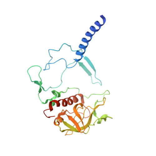
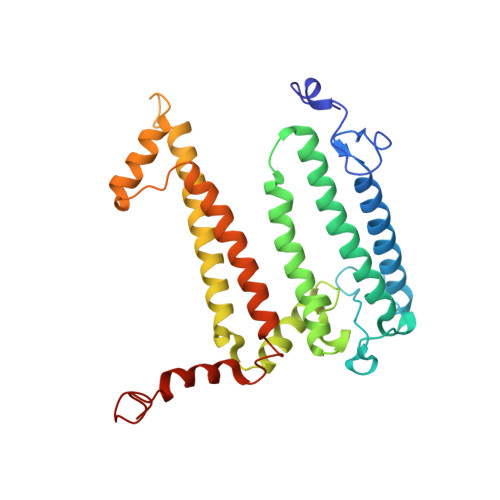
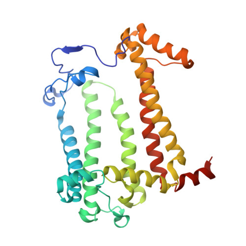
Structural and Spectroscopic Consequences of Hexa-Coordination of a Bacteriochlorophyll Cofactor in the Rhodobacter Sphaeroides Reaction Centre

Frolov, D.Marsh, M.Crouch, L.I.Fyfe, P.K.Robert, B.van Grondelle, R.Hadfield, A.T.Jones, M.R.

(2010) Biochemistry 49: 1882

  • DOI: https://doi.org/10.1021/bi901922t
  • Primary Citation of Related Structures:  
    2WX5

  • PubMed Abstract: 

    The structural and functional consequences of changing the coordination state of one of the bacteriochlorophyll (BChl) cofactors in the purple bacterial reaction center have been explored. A combination of steady state spectroscopy and X-ray crystallography was used to demonstrate that mutagenesis of residue 181 of the L-polypeptide from Phe to Arg (FL181R) causes the BChl at the accessory (B(B)) position on the so-called inactive cofactor branch to become hexacoordinated, with no significant changes to the structure of the surrounding protein. This change was accompanied by the appearance of a distinctive absorbance band at 631 nm in the room-temperature absorbance spectrum. The ligand donor was not the Arg side chain but rather an intervening water molecule, and contrary to expectations, the Mg of B(B) did not adopt a more in-plane geometry in response to hexacoordination. The mutation caused a disturbance to the detailed conformation of the BChl macrocycle that manifested in a number of subtle changes to the resonance Raman spectrum. Hexacoordination of B(B) produced a small increase in the lifetime of the excited electronic state of the primary donor bacteriochlorophylls (P*), indicating some disturbance to light-driven energy and/or electron transfer events on the time scale of a few picoseconds after light excitation. The B(B) bacteriochlorophyll returned to a pentacoordinated state in a double mutant where the FL181R mutation was combined with removal of the native axial ligand through mutation of His M182 to Leu. Experimental evidence of hexacoordinated bacteriochlorophylls in the literature on antenna proteins is considered, and possible reasons why hexacoordinated bacteriochlorophylls and chlorophylls appear to be avoided in photosynthetic proteins are discussed.


  • Organizational Affiliation

    Department of Physics and Astronomy, Free University of Amsterdam, De Boelelaan 1081, 1081 HV Amsterdam, The Netherlands.


Macromolecules
Find similar proteins by:  (by identity cutoff)  |  3D Structure
Entity ID: 1
MoleculeChains Sequence LengthOrganismDetailsImage
REACTION CENTER PROTEIN H CHAINA [auth H]260Cereibacter sphaeroidesMutation(s): 0 
Membrane Entity: Yes 
UniProt
Find proteins for Q3J170 (Cereibacter sphaeroides (strain ATCC 17023 / DSM 158 / JCM 6121 / CCUG 31486 / LMG 2827 / NBRC 12203 / NCIMB 8253 / ATH 2.4.1.))
Explore Q3J170 
Go to UniProtKB:  Q3J170
Entity Groups  
Sequence Clusters30% Identity50% Identity70% Identity90% Identity95% Identity100% Identity
UniProt GroupQ3J170
Sequence Annotations
Expand
  • Reference Sequence
Find similar proteins by:  (by identity cutoff)  |  3D Structure
Entity ID: 2
MoleculeChains Sequence LengthOrganismDetailsImage
REACTION CENTRE PROTEIN L CHAINB [auth L]281Cereibacter sphaeroidesMutation(s): 1 
Membrane Entity: Yes 
UniProt
Find proteins for Q3J1A5 (Cereibacter sphaeroides (strain ATCC 17023 / DSM 158 / JCM 6121 / CCUG 31486 / LMG 2827 / NBRC 12203 / NCIMB 8253 / ATH 2.4.1.))
Explore Q3J1A5 
Go to UniProtKB:  Q3J1A5
Entity Groups  
Sequence Clusters30% Identity50% Identity70% Identity90% Identity95% Identity100% Identity
UniProt GroupQ3J1A5
Sequence Annotations
Expand
  • Reference Sequence
Find similar proteins by:  (by identity cutoff)  |  3D Structure
Entity ID: 3
MoleculeChains Sequence LengthOrganismDetailsImage
REACTION CENTRE PROTEIN M CHAINC [auth M]307Cereibacter sphaeroidesMutation(s): 0 
Membrane Entity: Yes 
UniProt
Find proteins for Q3J1A6 (Cereibacter sphaeroides (strain ATCC 17023 / DSM 158 / JCM 6121 / CCUG 31486 / LMG 2827 / NBRC 12203 / NCIMB 8253 / ATH 2.4.1.))
Explore Q3J1A6 
Go to UniProtKB:  Q3J1A6
Entity Groups  
Sequence Clusters30% Identity50% Identity70% Identity90% Identity95% Identity100% Identity
UniProt GroupQ3J1A6
Sequence Annotations
Expand
  • Reference Sequence
Small Molecules
Ligands 10 Unique
IDChains Name / Formula / InChI Key2D Diagram3D Interactions
CDL
Query on CDL

Download Ideal Coordinates CCD File 
O [auth M]CARDIOLIPIN
C81 H156 O17 P2
XVTUQDWPJJBEHJ-KZCWQMDCSA-L
BCL
Query on BCL

Download Ideal Coordinates CCD File 
F [auth L],
G [auth L],
M,
N [auth M]
BACTERIOCHLOROPHYLL A
C55 H74 Mg N4 O6
DSJXIQQMORJERS-AGGZHOMASA-M
BPH
Query on BPH

Download Ideal Coordinates CCD File 
L,
X [auth M]
BACTERIOPHEOPHYTIN A
C55 H76 N4 O6
KWOZSBGNAHVCKG-SZQBJALDSA-N
U10
Query on U10

Download Ideal Coordinates CCD File 
Z [auth M]UBIQUINONE-10
C59 H90 O4
ACTIUHUUMQJHFO-UPTCCGCDSA-N
SPN
Query on SPN

Download Ideal Coordinates CCD File 
Y [auth M]SPEROIDENONE
C41 H70 O2
GWQAMGYOEYXWJF-YCDPMLDASA-N
LDA
Query on LDA

Download Ideal Coordinates CCD File 
D [auth H]
P [auth M]
Q [auth M]
R [auth M]
S [auth M]
D [auth H],
P [auth M],
Q [auth M],
R [auth M],
S [auth M],
T [auth M]
LAURYL DIMETHYLAMINE-N-OXIDE
C14 H31 N O
SYELZBGXAIXKHU-UHFFFAOYSA-N
HTO
Query on HTO

Download Ideal Coordinates CCD File 
J [auth L],
K [auth L],
W [auth M]
HEPTANE-1,2,3-TRIOL
C7 H16 O3
HXYCHJFUBNTKQR-RNFRBKRXSA-N
GOL
Query on GOL

Download Ideal Coordinates CCD File 
H [auth L],
I [auth L]
GLYCEROL
C3 H8 O3
PEDCQBHIVMGVHV-UHFFFAOYSA-N
FE
Query on FE

Download Ideal Coordinates CCD File 
V [auth M]FE (III) ION
Fe
VTLYFUHAOXGGBS-UHFFFAOYSA-N
NA
Query on NA

Download Ideal Coordinates CCD File 
E [auth H],
U [auth M]
SODIUM ION
Na
FKNQFGJONOIPTF-UHFFFAOYSA-N
Experimental Data & Validation

Experimental Data

  • Method: X-RAY DIFFRACTION
  • Resolution: 2.63 Å
  • R-Value Free: 0.279 
  • R-Value Work: 0.237 
  • R-Value Observed: 0.239 
  • Space Group: P 31 2 1
Unit Cell:
Length ( Å )Angle ( ˚ )
a = 140.18α = 90
b = 140.18β = 90
c = 185.401γ = 120
Software Package:
Software NamePurpose
REFMACrefinement
MOSFLMdata reduction
SCALAdata scaling
PHASERphasing

Structure Validation

View Full Validation Report


Ligand Structure Quality Assessment 


Entry History 

Deposition Data

Revision History  (Full details and data files)

  • Version 1.0: 2010-02-09
    Type: Initial release
  • Version 1.1: 2011-10-26
    Changes: Database references, Version format compliance
  • Version 1.2: 2017-12-13
    Changes: Database references, Structure summary
  • Version 1.3: 2023-12-20
    Changes: Data collection, Database references, Derived calculations, Other, Refinement description