2WSU

Galectin domain of porcine adenovirus type 4 NADC-1 isolate fibre


Experimental Data Snapshot

  • Method: X-RAY DIFFRACTION
  • Resolution: 1.90 Å
  • R-Value Free: 0.259 
  • R-Value Work: 0.206 
  • R-Value Observed: 0.208 

wwPDB Validation   3D Report Full Report


This is version 1.4 of the entry. See complete history


Literature

Crystallographic structure of porcine adenovirus type 4 fiber head and galectin domains.

Guardado-Calvo, P.Munoz, E.M.Llamas-Saiz, A.L.Fox, G.C.Kahn, R.Curiel, D.T.Glasgow, J.N.van Raaij, M.J.

(2010) J Virol 84: 10558-10568

  • DOI: https://doi.org/10.1128/JVI.00997-10
  • Primary Citation of Related Structures:  
    2WST, 2WSU, 2WSV, 2WT0, 2WT1, 2WT2

  • PubMed Abstract: 

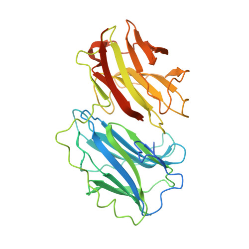
    Adenovirus isolate NADC-1, a strain of porcine adenovirus type 4, has a fiber containing an N-terminal virus attachment region, shaft and head domains, and a C-terminal galectin domain connected to the head by an RGD-containing sequence. The crystal structure of the head domain is similar to previously solved adenovirus fiber head domains, but specific residues for binding the coxsackievirus and adenovirus receptor (CAR), CD46, or sialic acid are not conserved. The structure of the galectin domain reveals an interaction interface between its two carbohydrate recognition domains, locating both sugar binding sites face to face. Sequence evidence suggests other tandem-repeat galectins have the same arrangement. We show that the galectin domain binds carbohydrates containing lactose and N-acetyl-lactosamine units, and we present structures of the galectin domain with lactose, N-acetyl-lactosamine, 3-aminopropyl-lacto-N-neotetraose, and 2-aminoethyl-tri(N-acetyl-lactosamine), confirming the domain as a bona fide galectin domain.


  • Organizational Affiliation

    Departamento de Bioquímica y Biología Molecular, Facultad de Farmacia, Universidad de Santiago de Compostela, E-15782 Santiago de Compostela, Spain.


Macromolecules
Find similar proteins by:  (by identity cutoff)  |  3D Structure
Entity ID: 1
MoleculeChains Sequence LengthOrganismDetailsImage
PUTATIVE FIBER PROTEIN
A, B, C, D
343Porcine adenovirus 4Mutation(s): 0 
UniProt
Find proteins for Q83467 (Porcine adenovirus B serotype 4)
Explore Q83467 
Go to UniProtKB:  Q83467
Entity Groups  
Sequence Clusters30% Identity50% Identity70% Identity90% Identity95% Identity100% Identity
UniProt GroupQ83467
Sequence Annotations
Expand
  • Reference Sequence
Small Molecules
Ligands 2 Unique
IDChains Name / Formula / InChI Key2D Diagram3D Interactions
GOL
Query on GOL

Download Ideal Coordinates CCD File 
E [auth A]
I [auth B]
J [auth B]
N [auth C]
O [auth C]
E [auth A],
I [auth B],
J [auth B],
N [auth C],
O [auth C],
S [auth D]
GLYCEROL
C3 H8 O3
PEDCQBHIVMGVHV-UHFFFAOYSA-N
NO3
Query on NO3

Download Ideal Coordinates CCD File 
F [auth A]
G [auth A]
H [auth A]
K [auth B]
L [auth B]
F [auth A],
G [auth A],
H [auth A],
K [auth B],
L [auth B],
M [auth B],
P [auth C],
Q [auth C],
R [auth C],
T [auth D],
U [auth D],
V [auth D]
NITRATE ION
N O3
NHNBFGGVMKEFGY-UHFFFAOYSA-N
Experimental Data & Validation

Experimental Data

  • Method: X-RAY DIFFRACTION
  • Resolution: 1.90 Å
  • R-Value Free: 0.259 
  • R-Value Work: 0.206 
  • R-Value Observed: 0.208 
  • Space Group: C 1 2 1
Unit Cell:
Length ( Å )Angle ( ˚ )
a = 167.691α = 90
b = 77.325β = 101.45
c = 94.111γ = 90
Software Package:
Software NamePurpose
REFMACrefinement
MOSFLMdata reduction
SCALAdata scaling
PHASERphasing

Structure Validation

View Full Validation Report


Entry History 

Deposition Data

Revision History  (Full details and data files)

  • Version 1.0: 2010-08-11
    Type: Initial release
  • Version 1.1: 2011-05-08
    Changes: Version format compliance
  • Version 1.2: 2011-07-13
    Changes: Version format compliance
  • Version 1.3: 2018-01-17
    Changes: Database references
  • Version 1.4: 2023-12-20
    Changes: Data collection, Database references, Derived calculations, Other, Refinement description