2WO8

MMP12 complex with a beta hydroxy carboxylic acid


Experimental Data Snapshot

  • Method: X-RAY DIFFRACTION
  • Resolution: 2.00 Å
  • R-Value Free: 0.246 
  • R-Value Work: 0.184 
  • R-Value Observed: 0.187 

wwPDB Validation   3D Report Full Report


Ligand Structure Quality Assessment 


This is version 1.2 of the entry. See complete history


Literature

The Identification of Beta-Hydroxy Carboxylic Acids as Selective Mmp-12 Inhibitors.

Holmes, I.P.Gaines, S.Watson, S.P.Lorthioir, O.Walker, A.Baddeley, S.J.Herbert, S.Egan, D.Convery, M.A.Singh, O.M.P.Gross, J.W.Strelow, J.M.Smith, R.H.Amour, A.J.Brown, D.Martin, S.L.

(2009) Bioorg Med Chem Lett 19: 5760

  • DOI: https://doi.org/10.1016/j.bmcl.2009.07.155
  • Primary Citation of Related Structures:  
    2WO8, 2WO9, 2WOA

  • PubMed Abstract: 

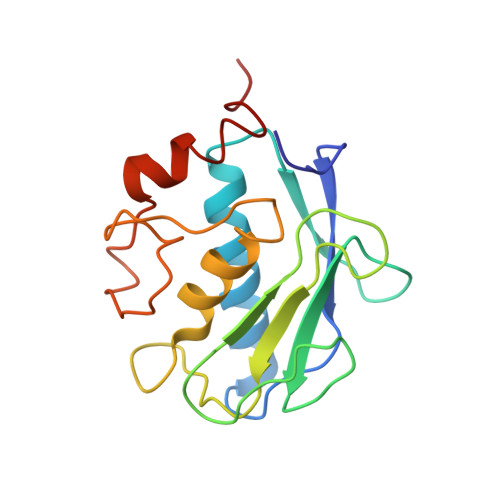
    A new class of selective MMP-12 inhibitors have been identified via high throughput screening. Crystallization with MMP-12 confirmed the mode of binding and allowed initial optimization to be carried out using classical structure based design.


  • Organizational Affiliation

    GSK Medicines Research Centre, Gunnels Wood Road, Stevenage, Hertfordshire SG1 2NY, United Kingdom.


Macromolecules
Find similar proteins by:  (by identity cutoff)  |  3D Structure
Entity ID: 1
MoleculeChains Sequence LengthOrganismDetailsImage
MACROPHAGE METALLOELASTASE
A, B, C, D
164Homo sapiensMutation(s): 0 
EC: 3.4.24.65
UniProt & NIH Common Fund Data Resources
Find proteins for P39900 (Homo sapiens)
Explore P39900 
Go to UniProtKB:  P39900
PHAROS:  P39900
GTEx:  ENSG00000262406 
Entity Groups  
Sequence Clusters30% Identity50% Identity70% Identity90% Identity95% Identity100% Identity
UniProt GroupP39900
Sequence Annotations
Expand
  • Reference Sequence
Small Molecules
Ligands 6 Unique
IDChains Name / Formula / InChI Key2D Diagram3D Interactions
023
Query on 023

Download Ideal Coordinates CCD File 
K [auth B],
V [auth D]
N^2^-[(2R)-2-{(1S)-1-[FORMYL(HYDROXY)AMINO]ETHYL}-5-PHENYLPENTANOYL]-N,3-DIMETHYL-L-VALINAMIDE
C21 H33 N3 O4
GHVMTHKJUAOZJP-CGTJXYLNSA-N
077
Query on 077

Download Ideal Coordinates CCD File 
H [auth A],
T [auth C]
(3S)-5-biphenyl-4-yl-3-hydroxypentanoic acid
C17 H18 O3
ILGSIIFHQGOKKV-INIZCTEOSA-N
SO4
Query on SO4

Download Ideal Coordinates CCD File 
CA [auth D],
I [auth A],
J [auth A],
P [auth B],
U [auth C]
SULFATE ION
O4 S
QAOWNCQODCNURD-UHFFFAOYSA-L
GOL
Query on GOL

Download Ideal Coordinates CCD File 
AA [auth D],
BA [auth D]
GLYCEROL
C3 H8 O3
PEDCQBHIVMGVHV-UHFFFAOYSA-N
ZN
Query on ZN

Download Ideal Coordinates CCD File 
E [auth A]
F [auth A]
L [auth B]
M [auth B]
Q [auth C]
E [auth A],
F [auth A],
L [auth B],
M [auth B],
Q [auth C],
R [auth C],
W [auth D],
X [auth D]
ZINC ION
Zn
PTFCDOFLOPIGGS-UHFFFAOYSA-N
CA
Query on CA

Download Ideal Coordinates CCD File 
G [auth A]
N [auth B]
O [auth B]
S [auth C]
Y [auth D]
G [auth A],
N [auth B],
O [auth B],
S [auth C],
Y [auth D],
Z [auth D]
CALCIUM ION
Ca
BHPQYMZQTOCNFJ-UHFFFAOYSA-N
Binding Affinity Annotations 
IDSourceBinding Affinity
077 Binding MOAD:  2WO8 IC50: 520 (nM) from 1 assay(s)
BindingDB:  2WO8 IC50: 520 (nM) from 1 assay(s)
Experimental Data & Validation

Experimental Data

  • Method: X-RAY DIFFRACTION
  • Resolution: 2.00 Å
  • R-Value Free: 0.246 
  • R-Value Work: 0.184 
  • R-Value Observed: 0.187 
  • Space Group: P 1
Unit Cell:
Length ( Å )Angle ( ˚ )
a = 46.043α = 66.47
b = 65.264β = 83.86
c = 67.808γ = 71.37
Software Package:
Software NamePurpose
REFMACrefinement
SCALEPACKdata scaling

Structure Validation

View Full Validation Report


Ligand Structure Quality Assessment 


Entry History 

Revision History  (Full details and data files)

  • Version 1.0: 2009-09-08
    Type: Initial release
  • Version 1.1: 2011-05-08
    Changes: Version format compliance
  • Version 1.2: 2011-07-13
    Changes: Version format compliance