Structural and Functional Characterization of a Triple Mutant Form of S100A7 Defective for Jab1 Binding.
West, N.R., Farnell, B., Murray, J.I., Hof, F., Watson, P.H., Boulanger, M.J.(2009) Protein Sci 18: 2615
- PubMed: 19844956
- DOI: https://doi.org/10.1002/pro.274
- Primary Citation of Related Structures:
2WND - PubMed Abstract:
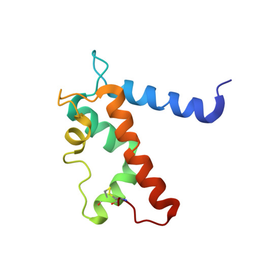
S100A7 (psoriasin) is a calcium- and zinc-binding protein implicated in breast cancer. We have shown previously that S100A7 enhances survival mechanisms in breast cells through an interaction with c-jun activation domain binding protein 1 (Jab1), and an engineered S100A7 triple mutant (Asp(56)Gly, Leu(78)Met, and Gln(88)Lys-S100A7(3)) ablates Jab1 binding. We extend these results to include defined breast cancer cell lines and demonstrate a disrupted S100A7(3)/Jab1 phenotype is maintained. To establish the basis for the abrogated Jab1 binding, we have recombinantly produced S100A7(3), demonstrated that it retains the ability to form an exceptionally thermostable dimer, and solved the three dimensional crystal structure to 1.6 A. Despite being positioned at the dimer interface, the Leu(78)Met mutation is easily accommodated and contributes to a methionine-rich pocket formed by Met(12), Met(15), and Met(34). In addition to altering the surface charge, the Gln(88)Lys mutation results in a nearby rotameric shift in Tyr(85), leading to a substantially reorganized surface cavity and may influence zinc binding. The final mutation of Asp(56) to Gly results in the largest structural perturbation shortening helix IV by one full turn. It is noteworthy that position 56 lies in one of two divergent clusters between S100A7 and the functionally distinct yet highly homologous S100A15. The structure of S100A7(3) provides a unique perspective from which to characterize the S100A7-Jab1 interaction and better understand the distinct functions between S100A7, and it is closely related paralog S100A15.
Organizational Affiliation:
Department of Biochemistry and Microbiology, University of Victoria, Victoria, British Columbia, Canada.