2WLG

Crystallographic analysis of the polysialic acid O-acetyltransferase OatWY


Experimental Data Snapshot

  • Method: X-RAY DIFFRACTION
  • Resolution: 1.90 Å
  • R-Value Free: 0.231 
  • R-Value Work: 0.193 
  • R-Value Observed: 0.195 

wwPDB Validation   3D Report Full Report


Ligand Structure Quality Assessment 


This is version 1.1 of the entry. See complete history


Literature

Structural and Kinetic Characterizations of the Polysialic Acid O-Acetyltransferase Oatwy from Neisseria Meningitidis.

Lee, H.J.Rakic, B.Gilbert, M.Wakarchuk, W.W.Withers, S.G.Strynadka, N.C.J.

(2009) J Biol Chem 284: 24501

  • DOI: https://doi.org/10.1074/jbc.M109.006049
  • Primary Citation of Related Structures:  
    2WLC, 2WLD, 2WLE, 2WLF, 2WLG

  • PubMed Abstract: 

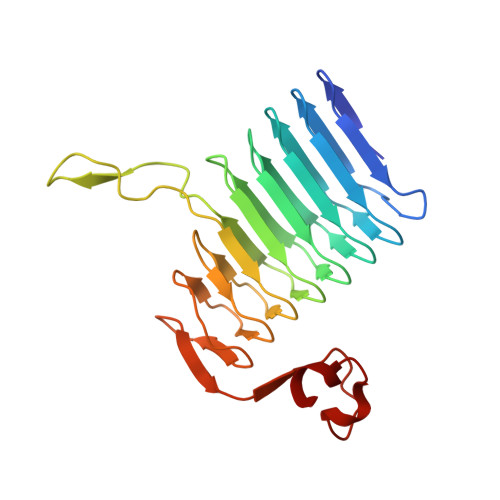
    The neuroinvasive pathogen Neisseria meningitidis has 13 capsular serogroups, but the majority of disease is caused by only 5 of these. Groups B, C, Y, and W-135 all display a polymeric sialic acid-containing capsule that provides a means for the bacteria to evade the immune response during infection by mimicking host sialic acid-containing cell surface structures. These capsules in serogroups C, Y, and W-135 can be further acetylated by a sialic acid-specific O-acetyltransferase, a modification that correlates with decreased immunoreactivity and increased virulence. In N. meningitidis serogroup Y, the O-acetylation reaction is catalyzed by the enzyme OatWY, which we show has clear specificity toward the serogroup Y capsule ([Glc-(alpha1-->4)-Sia](n)). To understand the underlying molecular basis of this process, we have performed crystallographic analysis of OatWY with bound substrate as well as determined kinetic parameters of the wild type enzyme and active site mutants. The structure of OatWY reveals an intimate homotrimer of left-handed beta-helix motifs that frame a deep active site cleft selective for the polysialic acid-bearing substrate. Within the active site, our structural, kinetic, and mutagenesis data support the role of two conserved residues in the catalytic mechanism (His-121 and Trp-145) and further highlight a significant movement of Tyr-171 that blocks the active site of the enzyme in its native form. Collectively, our results reveal the first structural features of a bacterial sialic acid O-acetyltransferase and provide significant new insight into its catalytic mechanism and specificity for the capsular polysaccharide of serogroup Y meningococci.


  • Organizational Affiliation

    Department of Biochemistry and Molecular Biology, University of British Columbia, Vancouver, British Columbia, Canada.


Macromolecules
Find similar proteins by:  (by identity cutoff)  |  3D Structure
Entity ID: 1
MoleculeChains Sequence LengthOrganismDetailsImage
POLYSIALIC ACID O-ACETYLTRANSFERASE
A, B, C
215Neisseria meningitidis serogroup YMutation(s): 1 
UniProt
Find proteins for Q93S40 (Neisseria meningitidis)
Explore Q93S40 
Go to UniProtKB:  Q93S40
Entity Groups  
Sequence Clusters30% Identity50% Identity70% Identity90% Identity95% Identity100% Identity
UniProt GroupQ93S40
Sequence Annotations
Expand
  • Reference Sequence
Small Molecules
Ligands 3 Unique
IDChains Name / Formula / InChI Key2D Diagram3D Interactions
SOP
Query on SOP

Download Ideal Coordinates CCD File 
D [auth A],
P [auth C]
[(2R,3S,4R,5R)-5-(6-AMINO-9H-PURIN-9-YL)-4-HYDROXY-3-(PHOSPHONOOXY)TETRAHYDROFURAN-2-YL]METHYL (3R)-3-HYDROXY-2,2-DIMETHYL-4-OXO-4-{[3-OXO-3-({2-[(2-OXOPROPYL)THIO]ETHYL}AMINO)PROPYL]AMINO}BUTYL DIHYDROGEN DIPHOSPHATE
C24 H40 N7 O17 P3 S
GAMKENBUYYJLCQ-NDZSKPAWSA-N
EDO
Query on EDO

Download Ideal Coordinates CCD File 
E [auth A]
F [auth A]
G [auth A]
H [auth A]
I [auth A]
E [auth A],
F [auth A],
G [auth A],
H [auth A],
I [auth A],
J [auth A],
K [auth A],
L [auth A],
N [auth B],
O [auth B],
S [auth C],
T [auth C],
U [auth C]
1,2-ETHANEDIOL
C2 H6 O2
LYCAIKOWRPUZTN-UHFFFAOYSA-N
ACT
Query on ACT

Download Ideal Coordinates CCD File 
M [auth B],
Q [auth C],
R [auth C]
ACETATE ION
C2 H3 O2
QTBSBXVTEAMEQO-UHFFFAOYSA-M
Experimental Data & Validation

Experimental Data

  • Method: X-RAY DIFFRACTION
  • Resolution: 1.90 Å
  • R-Value Free: 0.231 
  • R-Value Work: 0.193 
  • R-Value Observed: 0.195 
  • Space Group: P 2 2 2
Unit Cell:
Length ( Å )Angle ( ˚ )
a = 78.669α = 90
b = 94.393β = 90
c = 100.874γ = 90
Software Package:
Software NamePurpose
REFMACrefinement
HKLdata reduction
SCALEPACKdata scaling
PHASERphasing

Structure Validation

View Full Validation Report


Ligand Structure Quality Assessment 


Entry History 

Deposition Data

Revision History  (Full details and data files)

  • Version 1.0: 2009-06-30
    Type: Initial release
  • Version 1.1: 2011-07-13
    Changes: Advisory, Version format compliance