2WKS

Structure of Helicobacter pylori Type II Dehydroquinase with a new carbasugar-thiophene inhibitor.


Experimental Data Snapshot

  • Method: X-RAY DIFFRACTION
  • Resolution: 2.95 Å
  • R-Value Free: 0.236 
  • R-Value Work: 0.206 
  • R-Value Observed: 0.207 

wwPDB Validation   3D Report Full Report


Ligand Structure Quality Assessment 


This is version 1.5 of the entry. See complete history


Literature

Synthesis and biological evaluation of new nanomolar competitive inhibitors of Helicobacter pylori type II dehydroquinase. Structural details of the role of the aromatic moieties with essential residues.

Prazeres, V.F.Tizon, L.Otero, J.M.Guardado-Calvo, P.Llamas-Saiz, A.L.van Raaij, M.J.Castedo, L.Lamb, H.Hawkins, A.R.Gonzalez-Bello, C.

(2010) J Med Chem 53: 191-200

  • DOI: https://doi.org/10.1021/jm9010466
  • Primary Citation of Related Structures:  
    2WKS

  • PubMed Abstract: 

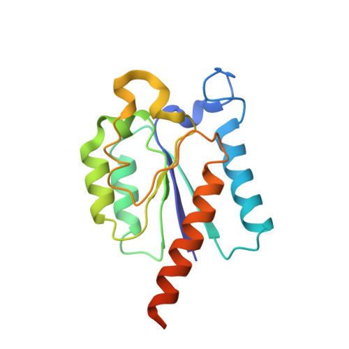
    The shikimic acid pathway is essential to many pathogens but absent in mammals. Enzymes in its pathway are therefore appropriate targets for the development of novel antibiotics. Dehydroquinase is the third enzyme of the pathway, catalyzing the reversible dehydratation of 3-dehydroquinic acid to form 3-dehydroshikimic acid. Here we present the synthesis of novel inhibitors with high affinity for Helicobacter pylori type II dehydroquinase and efficient inhibition characteristics. The structure of Helicobacter pylori type II dehydroquinase in complex with the most potent inhibitor shows that the aromatic functional group interacts with the catalytic Tyr22 by pi-stacking, expelling the Arg17 side chain, which is essential for catalysis, from the active site. The structure therefore explains the favorable properties of the inhibitor and will aid in design of improved antibiotics.


  • Organizational Affiliation

    Laboratorio de Quimica Organica (CSIC) y Departamento de Quimica Organica, Facultad de Quimica, Universidad de Santiago de Compostela, 15782 Santiago de Compostela, Spain.


Macromolecules
Find similar proteins by:  (by identity cutoff)  |  3D Structure
Entity ID: 1
MoleculeChains Sequence LengthOrganismDetailsImage
3-DEHYDROQUINATE DEHYDRATASE
A, B, C, D, E
A, B, C, D, E, F
167Helicobacter pyloriMutation(s): 0 
EC: 4.2.1.10
UniProt
Find proteins for Q48255 (Helicobacter pylori (strain ATCC 700392 / 26695))
Explore Q48255 
Go to UniProtKB:  Q48255
Entity Groups  
Sequence Clusters30% Identity50% Identity70% Identity90% Identity95% Identity100% Identity
UniProt GroupQ48255
Sequence Annotations
Expand
  • Reference Sequence
Small Molecules
Experimental Data & Validation

Experimental Data

  • Method: X-RAY DIFFRACTION
  • Resolution: 2.95 Å
  • R-Value Free: 0.236 
  • R-Value Work: 0.206 
  • R-Value Observed: 0.207 
  • Space Group: I 41
Unit Cell:
Length ( Å )Angle ( ˚ )
a = 157.85α = 90
b = 157.85β = 90
c = 99.78γ = 90
Software Package:
Software NamePurpose
REFMACrefinement
MOSFLMdata reduction
SCALAdata scaling
PHASERphasing

Structure Validation

View Full Validation Report


Ligand Structure Quality Assessment 


Entry History 

Revision History  (Full details and data files)

  • Version 1.0: 2009-11-24
    Type: Initial release
  • Version 1.1: 2011-06-30
    Changes: Version format compliance
  • Version 1.2: 2011-07-13
    Changes: Version format compliance
  • Version 1.3: 2018-02-14
    Changes: Database references
  • Version 1.4: 2019-07-24
    Changes: Data collection
  • Version 1.5: 2023-12-13
    Changes: Data collection, Database references, Derived calculations, Other, Refinement description