Insights Into the Biosynthesis of the Vibrio Cholerae Major Autoinducer Cai-1 from the Crystal Structure of the Plp-Dependent Enzyme Cqsa.
Jahan, N., Potter, J.A., Sheikh, M.A., Botting, C.H., Shirran, S.L., Westwood, N.J., Taylor, G.L.(2009) J Mol Biol 392: 763
- PubMed: 19631226
- DOI: https://doi.org/10.1016/j.jmb.2009.07.042
- Primary Citation of Related Structures:
2WK7, 2WK8, 2WK9, 2WKA - PubMed Abstract:
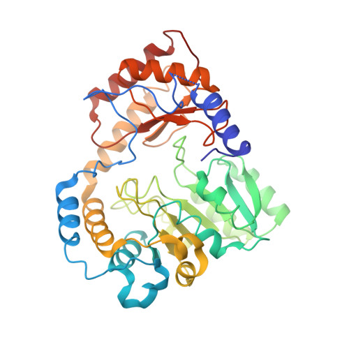
CqsA is an enzyme involved in the biosynthesis of cholerae autoinducer-1 (CAI-1), the major Vibrio cholerae autoinducer engaged in quorum sensing. The amino acid sequence of CqsA suggests that it belongs to the family of alpha-oxoamine synthases that catalyse the condensation of an amino acid to an acyl-CoA substrate. Here we present the apo- and PLP-bound crystal structures of CqsA and confirm that it shares structural homology with the dimeric alpha-oxoamine synthases, including a conserved PLP-binding site. The chemical structure of CAI-1 suggests that decanoyl-CoA may be one substrate of CqsA and that another substrate may be l-threonine or l-2-aminobutyric acid. A crystal structure of CqsA at 1.9-A resolution obtained in the presence of PLP and l-threonine reveals an external aldimine that has lost the l-threonine side chain. Similarly, a 1.9-A-resolution crystal structure of CqsA in the presence of PLP, l-threonine, and decanoyl-CoA shows a trapped external aldimine intermediate, suggesting that the condensation and decarboxylation steps have occurred, again with loss of the l-threonine side chain. It is suggested that this side-chain loss, an observation supported by mass spectrometry, is due to a retro-aldol reaction. Although no structural data have been obtained on CqsA using l-2-aminobutyric acid and decanoyl-CoA as substrates, mass spectrometry confirms the expected product of the enzyme reaction. It is proposed that a region of structure that is disordered in the apo structure is involved in the release of product. While not confirming if CqsA alone is able to synthesize CAI-1, these results suggest possible synthetic routes.
Organizational Affiliation:
Centre for Biomolecular Sciences, University of St. Andrews, UK.