2WGF

Crystal structure of Mycobacterium tuberculosis C171Q KasA variant


Experimental Data Snapshot

  • Method: X-RAY DIFFRACTION
  • Resolution: 2.15 Å
  • R-Value Free: 0.211 
  • R-Value Work: 0.166 
  • R-Value Observed: 0.168 

wwPDB Validation   3D Report Full Report


This is version 1.2 of the entry. See complete history


Literature

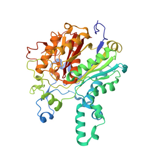
Crystal Structures of Mycobacterium Tuberculosis Kasa Show Mode of Action within Cell Wall Biosynthesis and its Inhibition by Thiolactomycin

Luckner, S.R.Machutta, C.A.Tonge, P.J.Kisker, C.

(2009) Structure 17: 1004

  • DOI: https://doi.org/10.1016/j.str.2009.04.012
  • Primary Citation of Related Structures:  
    2WGD, 2WGE, 2WGF, 2WGG

  • PubMed Abstract: 

    Mycobacteria have a unique cell wall consisting of mycolic acids, very-long-chain lipids that provide protection and allow the bacteria to persist within human macrophages. Inhibition of cell wall biosynthesis is fatal for the organism and a starting point for the discovery and development of novel antibiotics. We determined the crystal structures of KasA, a key enzyme involved in the biosynthesis of long-chain fatty acids, in its apo-form and bound to the natural product inhibitor thiolactomycin. Detailed insights into the interaction of the inhibitor with KasA and the identification of a polyethylene glycol molecule that mimics a fatty acid substrate of approximately 40 carbon atoms length, represent the first atomic view of a mycobacterial enzyme involved in the synthesis of long-chain fatty acids and provide a robust platform for the development of novel thiolactomycin analogs with high affinity for KasA.


  • Organizational Affiliation

    Rudolf Virchow Center for Experimental Biomedicine, Institute for Structural Biology, University of Würzburg, Würzburg, Germany.


Macromolecules
Find similar proteins by:  (by identity cutoff)  |  3D Structure
Entity ID: 1
MoleculeChains Sequence LengthOrganismDetailsImage
3-OXOACYL-[ACYL-CARRIER-PROTEIN] SYNTHASE 1
A, B, C, D, E
A, B, C, D, E, F, G, H
416Mycobacterium tuberculosisMutation(s): 1 
EC: 2.3.1.41
UniProt
Find proteins for P9WQD9 (Mycobacterium tuberculosis (strain ATCC 25618 / H37Rv))
Explore P9WQD9 
Go to UniProtKB:  P9WQD9
Entity Groups  
Sequence Clusters30% Identity50% Identity70% Identity90% Identity95% Identity100% Identity
UniProt GroupP9WQD9
Sequence Annotations
Expand
  • Reference Sequence
Small Molecules
Ligands 3 Unique
IDChains Name / Formula / InChI Key2D Diagram3D Interactions
PG4
Query on PG4

Download Ideal Coordinates CCD File 
J [auth A]
K [auth B]
L [auth B]
N [auth C]
P [auth D]
J [auth A],
K [auth B],
L [auth B],
N [auth C],
P [auth D],
S [auth E],
U [auth F]
TETRAETHYLENE GLYCOL
C8 H18 O5
UWHCKJMYHZGTIT-UHFFFAOYSA-N
PEG
Query on PEG

Download Ideal Coordinates CCD File 
Q [auth D]DI(HYDROXYETHYL)ETHER
C4 H10 O3
MTHSVFCYNBDYFN-UHFFFAOYSA-N
NA
Query on NA

Download Ideal Coordinates CCD File 
I [auth A]
M [auth B]
O [auth C]
R [auth D]
T [auth E]
I [auth A],
M [auth B],
O [auth C],
R [auth D],
T [auth E],
V [auth F],
W [auth G],
X [auth H]
SODIUM ION
Na
FKNQFGJONOIPTF-UHFFFAOYSA-N
Experimental Data & Validation

Experimental Data

  • Method: X-RAY DIFFRACTION
  • Resolution: 2.15 Å
  • R-Value Free: 0.211 
  • R-Value Work: 0.166 
  • R-Value Observed: 0.168 
  • Space Group: P 31
Unit Cell:
Length ( Å )Angle ( ˚ )
a = 151.505α = 90
b = 151.505β = 90
c = 147.839γ = 120
Software Package:
Software NamePurpose
PHENIXrefinement
MOSFLMdata reduction
TRUNCATEdata scaling
PHASERphasing

Structure Validation

View Full Validation Report


Entry History 

Deposition Data

Revision History  (Full details and data files)

  • Version 1.0: 2009-07-21
    Type: Initial release
  • Version 1.1: 2012-04-11
    Changes: Database references, Derived calculations, Non-polymer description, Other, Refinement description, Version format compliance
  • Version 1.2: 2023-12-13
    Changes: Data collection, Database references, Derived calculations, Other, Refinement description