2WD5

SMC hinge heterodimer (Mouse)


Experimental Data Snapshot

  • Method: X-RAY DIFFRACTION
  • Resolution: 2.70 Å
  • R-Value Free: 0.305 
  • R-Value Work: 0.225 
  • R-Value Observed: 0.229 

wwPDB Validation   3D Report Full Report


This is version 1.1 of the entry. See complete history


Literature

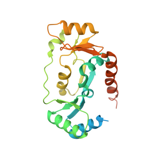
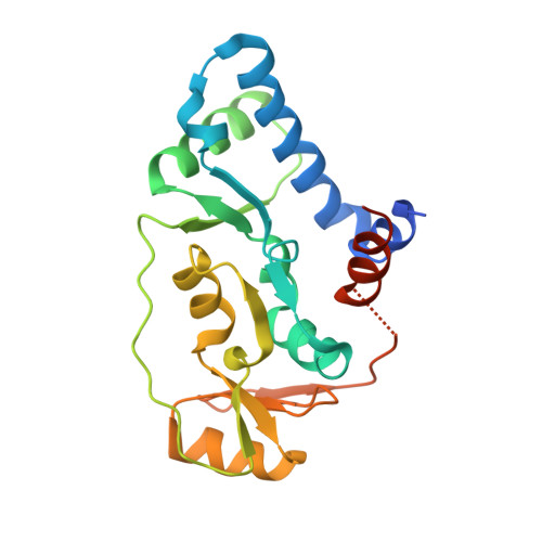
A Positively Charged Channel within the Smc1/Smc3 Hinge Required for Sister Chromatid Cohesion.

Kurze, A.Michie, K.A.Dixon, S.E.Mishra, A.Itoh, T.Khalid, S.Strmecki, L.Shirahige, K.Haering, C.H.Lowe, J.Nasmyth, K.

(2011) EMBO J 30: 364

  • DOI: https://doi.org/10.1038/emboj.2010.315
  • Primary Citation of Related Structures:  
    2WD5

  • PubMed Abstract: 

    Cohesin's structural maintenance of chromosome 1 (Smc1) and Smc3 are rod-shaped proteins with 50-nm long intra-molecular coiled-coil arms with a heterodimerization domain at one end and an ABC-like nucleotide-binding domain (NBD) at the other. Heterodimerization creates V-shaped molecules with a hinge at their centre. Inter-connection of NBDs by Scc1 creates a tripartite ring within which, it is proposed, sister DNAs are entrapped. To investigate whether cohesin's hinge functions as a possible DNA entry gate, we solved the crystal structure of the hinge from Mus musculus, which like its bacterial counterpart is characterized by a pseudo symmetric heterodimeric torus containing a small channel that is positively charged. Mutations in yeast Smc1 and Smc3 that together neutralize the channel's charge have little effect on dimerization or association with chromosomes, but are nevertheless lethal. Our finding that neutralization reduces acetylation of Smc3, which normally occurs during replication and is essential for cohesion, suggests that the positively charged channel is involved in a major conformational change during S phase.


  • Organizational Affiliation

    Department of Biochemistry, University of Oxford, Oxford, UK.


Macromolecules
Find similar proteins by:  (by identity cutoff)  |  3D Structure
Entity ID: 1
MoleculeChains Sequence LengthOrganismDetailsImage
STRUCTURAL MAINTENANCE OF CHROMOSOMES PROTEIN 1A233Mus musculusMutation(s): 0 
UniProt
Find proteins for Q9CU62 (Mus musculus)
Explore Q9CU62 
Go to UniProtKB:  Q9CU62
Entity Groups  
Sequence Clusters30% Identity50% Identity70% Identity90% Identity95% Identity100% Identity
UniProt GroupQ9CU62
Sequence Annotations
Expand
  • Reference Sequence
Find similar proteins by:  (by identity cutoff)  |  3D Structure
Entity ID: 2
MoleculeChains Sequence LengthOrganismDetailsImage
STRUCTURAL MAINTENANCE OF CHROMOSOMES PROTEIN 3213Mus musculusMutation(s): 0 
UniProt & NIH Common Fund Data Resources
Find proteins for Q9CW03 (Mus musculus)
Explore Q9CW03 
Go to UniProtKB:  Q9CW03
IMPC:  MGI:1339795
Entity Groups  
Sequence Clusters30% Identity50% Identity70% Identity90% Identity95% Identity100% Identity
UniProt GroupQ9CW03
Sequence Annotations
Expand
  • Reference Sequence
Experimental Data & Validation

Experimental Data

  • Method: X-RAY DIFFRACTION
  • Resolution: 2.70 Å
  • R-Value Free: 0.305 
  • R-Value Work: 0.225 
  • R-Value Observed: 0.229 
  • Space Group: P 1 21 1
Unit Cell:
Length ( Å )Angle ( ˚ )
a = 52.285α = 90
b = 61.867β = 105.04
c = 77.046γ = 90
Software Package:
Software NamePurpose
PHENIXrefinement
MOSFLMdata reduction
SCALAdata scaling
SHARPphasing

Structure Validation

View Full Validation Report


Entry History 

Deposition Data

Revision History  (Full details and data files)

  • Version 1.0: 2010-08-11
    Type: Initial release
  • Version 1.1: 2012-10-10
    Changes: Database references, Structure summary, Version format compliance