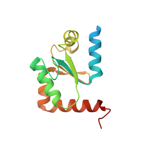
2WCI

Structure of E. coli monothiol glutaredoxin GRX4 homodimer


Experimental Data Snapshot

  • Method: X-RAY DIFFRACTION
  • Resolution: 1.90 Å
  • R-Value Free: 0.234 
  • R-Value Work: 0.183 
  • R-Value Observed: 0.186 

wwPDB Validation   3D Report Full Report


Ligand Structure Quality Assessment 


This is version 1.1 of the entry. See complete history


Literature

Structural Basis for Delivery of the Intact [Fe2S2] Cluster by Monothiol Glutaredoxin.

Iwema, T.Picciocchi, A.Traore, D.A.K.Ferrer, J.-L.Chauvat, F.Jacquamet, L.

(2009) Biochemistry 48: 6041

  • DOI: https://doi.org/10.1021/bi900440m
  • Primary Citation of Related Structures:  
    2WCI

  • PubMed Abstract: 

    Glutaredoxins (GRX) are redox proteins which use glutathione as a cofactor and are divided into two classes, monothiol and dithiol. In each class, several GRX have been shown to form [Fe2S2] cluster coordinating homodimers. The dithiol GRX homodimer is proposed to serve as a sequestration form and its iron-sulfur cluster as an oxidative stress sensor. In contrast, the monothiol GRX homodimer has been suggested to act as a scaffold for [Fe2S2] cluster delivery. We present here the structure of a monothiol GRX homodimer (Escherichia coli GRX4) coordinating a [Fe2S2] cluster that reveals the structural basis of intact iron-sulfur cluster delivery.


  • Organizational Affiliation

    Laboratoire de Cristallographie et Cristallogenèse des Protéines, Institut de Biologie Structurale Jean-Pierre Ebel, 5075 CEA, CNRS, Universite Joseph Fourier, 41 rue Jules Horowitz, F-38027 Grenoble, France. thomas.iwema@ibs.fr


Macromolecules
Find similar proteins by:  (by identity cutoff)  |  3D Structure
Entity ID: 1
MoleculeChains Sequence LengthOrganismDetailsImage
GLUTAREDOXIN-4
A, B
135Escherichia coliMutation(s): 0 
UniProt
Find proteins for P0AC69 (Escherichia coli (strain K12))
Explore P0AC69 
Go to UniProtKB:  P0AC69
Entity Groups  
Sequence Clusters30% Identity50% Identity70% Identity90% Identity95% Identity100% Identity
UniProt GroupP0AC69
Sequence Annotations
Expand
  • Reference Sequence
Experimental Data & Validation

Experimental Data

  • Method: X-RAY DIFFRACTION
  • Resolution: 1.90 Å
  • R-Value Free: 0.234 
  • R-Value Work: 0.183 
  • R-Value Observed: 0.186 
  • Space Group: P 43 21 2
Unit Cell:
Length ( Å )Angle ( ˚ )
a = 94.3α = 90
b = 94.3β = 90
c = 62.66γ = 90
Software Package:
Software NamePurpose
REFMACrefinement
XDSdata reduction
XSCALEdata scaling
SHELXphasing

Structure Validation

View Full Validation Report


Ligand Structure Quality Assessment 


Entry History 

Deposition Data

Revision History  (Full details and data files)

  • Version 1.0: 2009-06-23
    Type: Initial release
  • Version 1.1: 2012-02-29
    Changes: Advisory, Atomic model, Database references, Derived calculations, Non-polymer description, Other, Structure summary