2WAJ

Crystal structure of human Jnk3 complexed with a 1-aryl-3,4- dihydroisoquinoline inhibitor


Experimental Data Snapshot

  • Method: X-RAY DIFFRACTION
  • Resolution: 2.40 Å
  • R-Value Free: 0.264 
  • R-Value Work: 0.183 
  • R-Value Observed: 0.187 

wwPDB Validation   3D Report Full Report


Ligand Structure Quality Assessment 


This is version 1.2 of the entry. See complete history


Literature

1-Aryl-3,4-Dihydroisoquinoline Inhibitors of Jnk3.

Christopher, J.A.Atkinson, F.L.Bax, B.D.Brown, M.J.Champigny, A.C.Chuang, T.T.Jones, E.J.Mosley, J.E.Musgrave, J.R.

(2009) Bioorg Med Chem Lett 19: 2230

  • DOI: https://doi.org/10.1016/j.bmcl.2009.02.098
  • Primary Citation of Related Structures:  
    2WAJ

  • PubMed Abstract: 

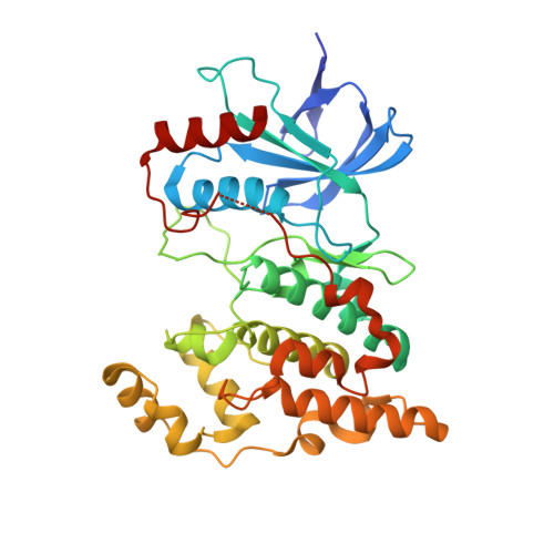
    A series of 1-aryl-3,4-dihydroisoquinoline inhibitors of JNK3 are described. Compounds 20 and 24 are the most potent inhibitors (pIC50 7.3 and 6.9, respectively in a radiometric filter binding assay), with 10- and 1000-fold selectivity over JNK2 and JNK1, respectively, and selectivity within the wider mitogen-activated protein kinase (MAPK) family against p38alpha and ERK2. X-ray crystallography of 16 reveals a highly unusual binding mode where an H-bond acceptor interaction with the hinge region is made by a chloro substituent.


  • Organizational Affiliation

    GlaxoSmithKline R&D, Medicines Research Centre, Stevenage, Hertfordshire, UK. john@christopher257.fsnet.co.uk


Macromolecules
Find similar proteins by:  (by identity cutoff)  |  3D Structure
Entity ID: 1
MoleculeChains Sequence LengthOrganismDetailsImage
MITOGEN-ACTIVATED PROTEIN KINASE 10364Homo sapiensMutation(s): 0 
EC: 2.7.11.24
UniProt & NIH Common Fund Data Resources
Find proteins for P53779 (Homo sapiens)
Explore P53779 
Go to UniProtKB:  P53779
PHAROS:  P53779
GTEx:  ENSG00000109339 
Entity Groups  
Sequence Clusters30% Identity50% Identity70% Identity90% Identity95% Identity100% Identity
UniProt GroupP53779
Sequence Annotations
Expand
  • Reference Sequence
Small Molecules
Ligands 1 Unique
IDChains Name / Formula / InChI Key2D Diagram3D Interactions
SNB
Query on SNB

Download Ideal Coordinates CCD File 
B [auth A]1-(3-BROMOPHENYL)-7-CHLORO-6-METHOXY-3,4-DIHYDROISOQUINOLINE
C16 H13 Br Cl N O
IKGXHBGCVQTQBH-UHFFFAOYSA-N
Binding Affinity Annotations 
IDSourceBinding Affinity
SNB PDBBind:  2WAJ IC50: 398 (nM) from 1 assay(s)
Binding MOAD:  2WAJ IC50: 398 (nM) from 1 assay(s)
BindingDB:  2WAJ IC50: 398.11 (nM) from 1 assay(s)
Experimental Data & Validation

Experimental Data

  • Method: X-RAY DIFFRACTION
  • Resolution: 2.40 Å
  • R-Value Free: 0.264 
  • R-Value Work: 0.183 
  • R-Value Observed: 0.187 
  • Space Group: P 21 21 21
Unit Cell:
Length ( Å )Angle ( ˚ )
a = 51.659α = 90
b = 71.339β = 90
c = 107.541γ = 90
Software Package:
Software NamePurpose
REFMACrefinement
MOSFLMdata reduction
SCALAdata scaling
REFMACphasing

Structure Validation

View Full Validation Report


Ligand Structure Quality Assessment 


Entry History 

Deposition Data

Revision History  (Full details and data files)

  • Version 1.0: 2009-03-31
    Type: Initial release
  • Version 1.1: 2011-07-13
    Changes: Advisory, Version format compliance
  • Version 1.2: 2023-12-13
    Changes: Data collection, Database references, Derived calculations, Other, Refinement description