2W9R

Structural basis of N-end rule substrate recognition in Escherichia coli by the ClpAP adaptor protein ClpS


Experimental Data Snapshot

  • Method: X-RAY DIFFRACTION
  • Resolution: 1.70 Å
  • R-Value Free: 0.254 
  • R-Value Work: 0.226 
  • R-Value Observed: 0.228 

wwPDB Validation   3D Report Full Report


This is version 1.2 of the entry. See complete history

Re-refinement Note

A newer entry is available that reflects an alternative modeling of the original data: 3O2H


Literature

Structural Basis of N-End Rule Substrate Recognition in Escherichia Coli by the Clpap Adaptor Protein Clps.

Schuenemann, V.J.Kralik, S.M.Albrecht, R.Spall, S.K.Truscott, K.N.Dougan, D.A.Zeth, K.

(2009) EMBO Rep 10: 508

  • DOI: https://doi.org/10.1038/embor.2009.62
  • Primary Citation of Related Structures:  
    2W9R, 2WA8, 2WA9

  • PubMed Abstract: 

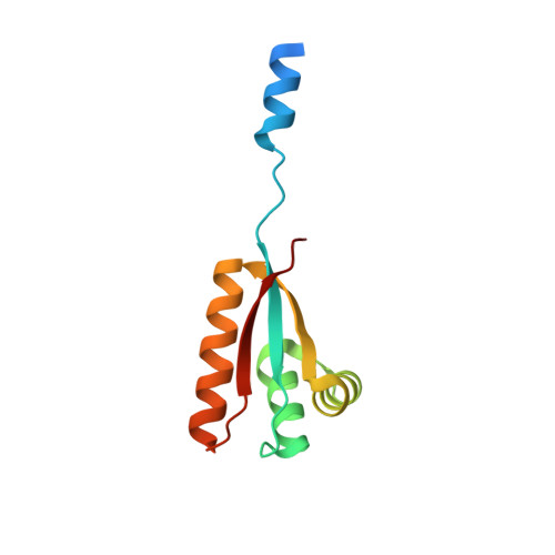
    In Escherichia coli, the ClpAP protease, together with the adaptor protein ClpS, is responsible for the degradation of proteins bearing an amino-terminal destabilizing amino acid (N-degron). Here, we determined the three-dimensional structures of ClpS in complex with three peptides, each having a different destabilizing residue--Leu, Phe or Trp--at its N terminus. All peptides, regardless of the identity of their N-terminal residue, are bound in a surface pocket on ClpS in a stereo-specific manner. Several highly conserved residues in this binding pocket interact directly with the backbone of the N-degron peptide and hence are crucial for the binding of all N-degrons. By contrast, two hydrophobic residues define the volume of the binding pocket and influence the specificity of ClpS. Taken together, our data suggest that ClpS has been optimized for the binding and delivery of N-degrons containing an N-terminal Phe or Leu.


  • Organizational Affiliation

    Department of Protein Evolution, Max Planck Institute for Developmental Biology, Spemannstrasse 35, Tübingen D-72076, Germany.


Macromolecules
Find similar proteins by:  (by identity cutoff)  |  3D Structure
Entity ID: 1
MoleculeChains Sequence LengthOrganismDetailsImage
ATP-DEPENDENT CLP PROTEASE ADAPTER PROTEIN CLPS108Escherichia coli K-12Mutation(s): 0 
UniProt
Find proteins for P0A8Q6 (Escherichia coli (strain K12))
Explore P0A8Q6 
Go to UniProtKB:  P0A8Q6
Entity Groups  
Sequence Clusters30% Identity50% Identity70% Identity90% Identity95% Identity100% Identity
UniProt GroupP0A8Q6
Sequence Annotations
Expand
  • Reference Sequence

Find similar proteins by:  Sequence   |   3D Structure  

Entity ID: 2
MoleculeChains Sequence LengthOrganismDetailsImage
DNA PROTECTION DURING STARVATION PROTEIN11Escherichia coli K-12Mutation(s): 0 
UniProt
Find proteins for P0ABT2 (Escherichia coli (strain K12))
Explore P0ABT2 
Go to UniProtKB:  P0ABT2
Entity Groups  
Sequence Clusters30% Identity50% Identity70% Identity90% Identity95% Identity100% Identity
UniProt GroupP0ABT2
Sequence Annotations
Expand
  • Reference Sequence
Experimental Data & Validation

Experimental Data

  • Method: X-RAY DIFFRACTION
  • Resolution: 1.70 Å
  • R-Value Free: 0.254 
  • R-Value Work: 0.226 
  • R-Value Observed: 0.228 
  • Space Group: P 1
Unit Cell:
Length ( Å )Angle ( ˚ )
a = 28.08α = 97.44
b = 28.23β = 106.45
c = 38.91γ = 92.39
Software Package:
Software NamePurpose
REFMACrefinement
XDSdata reduction
XDSdata scaling

Structure Validation

View Full Validation Report


Entry History 

Deposition Data

Revision History  (Full details and data files)

  • Version 1.0: 2009-04-28
    Type: Initial release
  • Version 1.1: 2011-05-08
    Changes: Version format compliance
  • Version 1.2: 2011-07-13
    Changes: Version format compliance