2W9G

Wild-type Staphylococcus aureus DHFR in complex with NADPH and trimethoprim


Experimental Data Snapshot

  • Method: X-RAY DIFFRACTION
  • Resolution: 1.95 Å
  • R-Value Free: 0.287 
  • R-Value Work: 0.211 
  • R-Value Observed: 0.214 

wwPDB Validation   3D Report Full Report


Ligand Structure Quality Assessment 


This is version 1.2 of the entry. See complete history


Literature

Structural Comparison of Chromosomal and Exogenous Dihydrofolate Reductase from Staphylococcus Aureus in Complex with the Potent Inhibitor Trimethoprim.

Heaslet, H.Harris, M.Fahnoe, K.Sarver, R.Putz, H.Chang, J.Subramanian, C.Barreiro, G.Miller, J.R.

(2009) Proteins 76: 706

  • DOI: https://doi.org/10.1002/prot.22383
  • Primary Citation of Related Structures:  
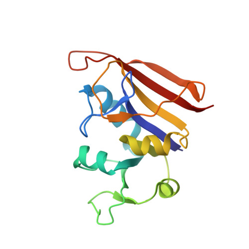
    2W9G, 2W9H, 2W9S, 2W9T

  • PubMed Abstract: 

    Dihydrofolate reductase (DHFR) is the enzyme responsible for the NADPH-dependent reduction of 5,6-dihydrofolate to 5,6,7,8-tetrahydrofolate, an essential cofactor in the synthesis of purines, thymidylate, methionine, and other key metabolites. Because of its importance in multiple cellular functions, DHFR has been the subject of much research targeting the enzyme with anticancer, antibacterial, and antimicrobial agents. Clinically used compounds targeting DHFR include methotrexate for the treatment of cancer and diaminopyrimidines (DAPs) such as trimethoprim (TMP) for the treatment of bacterial infections. DAP inhibitors of DHFR have been used clinically for >30 years and resistance to these agents has become widespread. Methicillin-resistant Staphylococcus aureus (MRSA), the causative agent of many serious nosocomial and community acquired infections, and other gram-positive organisms can show resistance to DAPs through mutation of the chromosomal gene or acquisition of an alternative DHFR termed "S1 DHFR." To develop new therapies for health threats such as MRSA, it is important to understand the molecular basis of DAP resistance. Here, we report the crystal structure of the wild-type chromosomal DHFR from S. aureus in complex with NADPH and TMP. We have also solved the structure of the exogenous, TMP resistant S1 DHFR, apo and in complex with TMP. The structural and thermodynamic data point to important molecular differences between the two enzymes that lead to dramatically reduced affinity of DAPs to S1 DHFR. These differences in enzyme binding affinity translate into reduced antibacterial activity against strains of S. aureus that express S1 DHFR.


  • Organizational Affiliation

    Lead Development Technologies, Pfizer Global Research and Development, Groton, Connecticut 06340, USA. holly.h.soutter@pfizer.com


Macromolecules
Find similar proteins by:  (by identity cutoff)  |  3D Structure
Entity ID: 1
MoleculeChains Sequence LengthOrganismDetailsImage
DIHYDROFOLATE REDUCTASE159Staphylococcus aureusMutation(s): 0 
EC: 1.5.1.3
UniProt
Find proteins for P0A017 (Staphylococcus aureus)
Explore P0A017 
Go to UniProtKB:  P0A017
Entity Groups  
Sequence Clusters30% Identity50% Identity70% Identity90% Identity95% Identity100% Identity
UniProt GroupP0A017
Sequence Annotations
Expand
  • Reference Sequence
Small Molecules
Ligands 2 Unique
IDChains Name / Formula / InChI Key2D Diagram3D Interactions
NDP
Query on NDP

Download Ideal Coordinates CCD File 
C [auth A]NADPH DIHYDRO-NICOTINAMIDE-ADENINE-DINUCLEOTIDE PHOSPHATE
C21 H30 N7 O17 P3
ACFIXJIJDZMPPO-NNYOXOHSSA-N
TOP
Query on TOP

Download Ideal Coordinates CCD File 
B [auth A]TRIMETHOPRIM
C14 H18 N4 O3
IEDVJHCEMCRBQM-UHFFFAOYSA-N
Binding Affinity Annotations 
IDSourceBinding Affinity
TOP BindingDB:  2W9G Ki: min: 1.2, max: 5500 (nM) from 7 assay(s)
IC50: min: 7, max: 4.50e+4 (nM) from 5 assay(s)
Binding MOAD:  2W9G Kd: 0.97 (nM) from 1 assay(s)
NDP Binding MOAD:  2W9G Kd: 8.1 (nM) from 1 assay(s)
Experimental Data & Validation

Experimental Data

  • Method: X-RAY DIFFRACTION
  • Resolution: 1.95 Å
  • R-Value Free: 0.287 
  • R-Value Work: 0.211 
  • R-Value Observed: 0.214 
  • Space Group: P 61 2 2
Unit Cell:
Length ( Å )Angle ( ˚ )
a = 79.287α = 90
b = 79.287β = 90
c = 106.254γ = 120
Software Package:
Software NamePurpose
REFMACrefinement
HKL-2000data reduction
SCALEPACKdata scaling
AMoREphasing

Structure Validation

View Full Validation Report


Ligand Structure Quality Assessment 


Entry History 

Deposition Data

Revision History  (Full details and data files)

  • Version 1.0: 2009-03-24
    Type: Initial release
  • Version 1.1: 2011-05-08
    Changes: Version format compliance
  • Version 1.2: 2011-07-13
    Changes: Version format compliance