2W5I

RNASE A-AP3A COMPLEX


Experimental Data Snapshot

  • Method: X-RAY DIFFRACTION
  • Resolution: 2.40 Å
  • R-Value Free: 0.273 
  • R-Value Work: 0.190 
  • R-Value Observed: 0.198 

wwPDB Validation   3D Report Full Report


Ligand Structure Quality Assessment 


This is version 1.3 of the entry. See complete history


Literature

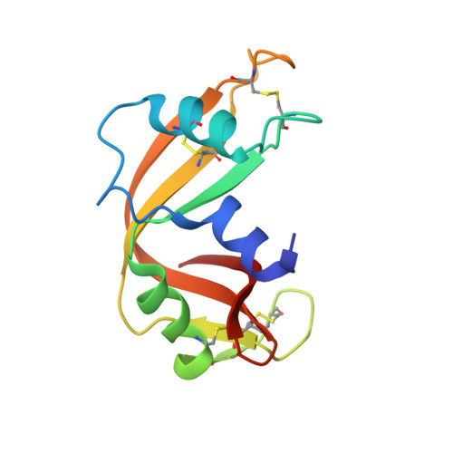
Influence of Naturally-Occurring 5'-Pyrophosphate-Linked Substituents on the Binding of Adenylic Inhibitors to Ribonuclease A: An X-Ray Crystallographic Study.

Holloway, D.E.Chavali, G.B.Leonidas, D.D.Baker, M.D.Acharya, K.R.

(2009) Biopolymers 91: 995

  • DOI: https://doi.org/10.1002/bip.21158
  • Primary Citation of Related Structures:  
    2W5G, 2W5I, 2W5K, 2W5L, 2W5M

  • PubMed Abstract: 

    Ribonuclease A is the archetype of a functionally diverse superfamily of vertebrate-specific ribonucleases. Inhibitors of its action have potential use in the elucidation of the in vivo roles of these enzymes and in the treatment of pathologies associated therewith. Derivatives of adenosine 5'-pyrophosphate are the most potent nucleotide-based inhibitors known. Here, we use X-ray crystallography to visualize the binding of four naturally-occurring derivatives that contain 5'-pyrophosphate-linked extensions. 5'-ATP binds with the adenine occupying the B(2) subsite in the manner of an RNA substrate but with the gamma-phosphate at the P(1) subsite. Diadenosine triphosphate (Ap(3)A) binds with the adenine in syn conformation, the beta-phosphate as the principal P(1) subsite ligand and without order beyond the gamma-phosphate. NADPH and NADP(+) bind with the adenine stacked against an alternative rotamer of His119, the 2'-phosphate at the P(1) subsite, and without order beyond the 5'-alpha-phosphate. We also present the structure of the complex formed with pyrophosphate ion. The structural data enable existing kinetic data on the binding of these compounds to a variety of ribonucleases to be rationalized and suggest that as the complexity of the 5'-linked extension increases, the need to avoid unfavorable contacts places limitations on the number of possible binding modes.


  • Organizational Affiliation

    Department of Biology and Biochemistry, University of Bath, Claverton Down, Bath BA2 7AY, UK.


Macromolecules
Find similar proteins by:  (by identity cutoff)  |  3D Structure
Entity ID: 1
MoleculeChains Sequence LengthOrganismDetailsImage
RIBONUCLEASE PANCREATIC
A, B
124Bos taurusMutation(s): 0 
EC: 3.1.27.5
UniProt
Find proteins for P61823 (Bos taurus)
Explore P61823 
Go to UniProtKB:  P61823
Entity Groups  
Sequence Clusters30% Identity50% Identity70% Identity90% Identity95% Identity100% Identity
UniProt GroupP61823
Sequence Annotations
Expand
  • Reference Sequence
Small Molecules
Ligands 1 Unique
IDChains Name / Formula / InChI Key2D Diagram3D Interactions
ATP
Query on ATP

Download Ideal Coordinates CCD File 
C [auth A],
D [auth B]
ADENOSINE-5'-TRIPHOSPHATE
C10 H16 N5 O13 P3
ZKHQWZAMYRWXGA-KQYNXXCUSA-N
Binding Affinity Annotations 
IDSourceBinding Affinity
ATP Binding MOAD:  2W5I Ki: 860 (nM) from 1 assay(s)
PDBBind:  2W5I Ki: 2.90e+4 (nM) from 1 assay(s)
Experimental Data & Validation

Experimental Data

  • Method: X-RAY DIFFRACTION
  • Resolution: 2.40 Å
  • R-Value Free: 0.273 
  • R-Value Work: 0.190 
  • R-Value Observed: 0.198 
  • Space Group: C 1 2 1
Unit Cell:
Length ( Å )Angle ( ˚ )
a = 101.96α = 90
b = 33.402β = 91.05
c = 75.703γ = 90
Software Package:
Software NamePurpose
REFMACrefinement
DENZOdata reduction
SCALEPACKdata scaling
AMoREphasing

Structure Validation

View Full Validation Report


Ligand Structure Quality Assessment 


Entry History 

Deposition Data

Revision History  (Full details and data files)

  • Version 1.0: 2009-02-17
    Type: Initial release
  • Version 1.1: 2011-05-08
    Changes: Version format compliance
  • Version 1.2: 2011-07-13
    Changes: Version format compliance
  • Version 1.3: 2023-12-13
    Changes: Data collection, Database references, Derived calculations, Other, Refinement description