Hise11 and Hisf8 Provide Bis-Histidyl Heme Hexa-Coordination in the Globin Domain of Geobacter Sulfurreducens Globin-Coupled Sensor.
Pesce, A., Thijs, L., Nardini, M., Desmet, F., Sisinni, L., Gourlay, L., Bolli, A., Coletta, M., Van Doorslaer, S., Wan, X., Alam, M., Ascenzi, P., Moens, L., Bolognesi, M., Dewilde, S.(2009) J Mol Biol 386: 246
- PubMed: 19109973
- DOI: https://doi.org/10.1016/j.jmb.2008.12.023
- Primary Citation of Related Structures:
2W31 - PubMed Abstract:
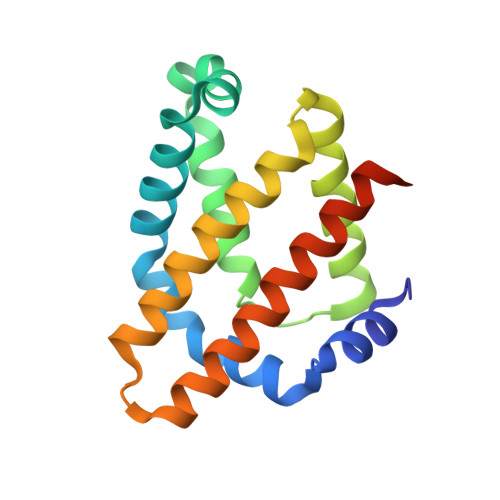
Among heme-based sensors, recent phylogenomic and sequence analyses have identified 34 globin coupled sensors (GCS), to which an aerotactic or gene-regulating function has been tentatively ascribed. Here, the structural and biochemical characterization of the globin domain of the GCS from Geobacter sulfurreducens (GsGCS(162)) is reported. A combination of X-ray crystallography (crystal structure at 1.5 A resolution), UV-vis and resonance Raman spectroscopy reveals the ferric GsGCS(162) as an example of bis-histidyl hexa-coordinated GCS. In contrast to the known hexa-coordinated globins, the distal heme-coordination in ferric GsGCS(162) is provided by a His residue unexpectedly located at the E11 topological site. Furthermore, UV-vis and resonance Raman spectroscopy indicated that ferrous deoxygenated GsGCS(162) is a penta-/hexa-coordinated mixture, and the heme hexa-to-penta-coordination transition does not represent a rate-limiting step for carbonylation kinetics. Lastly, electron paramagnetic resonance indicates that ferrous nitrosylated GsGCS(162) is a penta-coordinated species, where the proximal HisF8-Fe bond is severed.
Organizational Affiliation:
Department of Physics, CNISM and Center for Excellence in Biomedical Research, University of Genova, Via Dodecaneso, 33, I-16146 Genova, Italy.