Functional Proteomic and Structural Insights Into Molecular Recognition in the Nitrilase Family Enzymes.
Barglow, K.T., Saikatendu, K.S., Bracey, M.H., Huey, R., Morris, G.M., Olson, A.J., Stevens, R.C., Cravatt, B.F.(2008) Biochemistry 47: 13514
- PubMed: 19053248
- DOI: https://doi.org/10.1021/bi801786y
- Primary Citation of Related Structures:
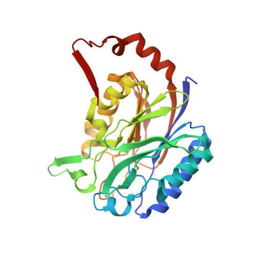
2W1V - PubMed Abstract:
Nitrilases are a large and diverse family of nonpeptidic C-N hydrolases. The mammalian genome encodes eight nitrilase enzymes, several of which remain poorly characterized. Prominent among these are nitrilase-1 (Nit1) and nitrilase-2 (Nit2), which, despite having been shown to exert effects on cell growth and possibly serving as tumor suppressor genes, are without known substrates or selective inhibitors. In previous studies, we identified several nitrilases, including Nit1 and Nit2, as targets for dipeptide-chloroacetamide activity-based proteomics probes. Here, we have used these probes, in combination with high-resolution crystallography and molecular modeling, to systematically map the active site of Nit2 and identify residues involved in molecular recognition. We report the 1.4 A crystal structure of mouse Nit2 and use this structure to identify residues that discriminate probe labeling between the Nit1 and Nit2 enzymes. Interestingly, some of these residues are conserved across all vertebrate Nit2 enzymes and, conversely, not found in any vertebrate Nit1 enzymes, suggesting that they are key discriminators of molecular recognition between these otherwise highly homologous enzymes. Our findings thus point to a limited set of active site residues that establish distinct patterns of molecular recognition among nitrilases and provide chemical probes to selectively perturb the function of these enzymes in biological systems.
Organizational Affiliation:
The Skaggs Institute for Chemical Biology and Department of Chemical Physiology, The Scripps Research Institute, 10550 North Torrey Pines Road, La Jolla, California 92037, USA.