2W10

Mona SH3C in complex


Experimental Data Snapshot

  • Method: X-RAY DIFFRACTION
  • Resolution: 1.90 Å
  • R-Value Free: 0.231 
  • R-Value Work: 0.161 
  • R-Value Observed: 0.164 

wwPDB Validation   3D Report Full Report


This is version 1.3 of the entry. See complete history


Literature

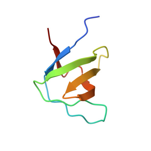
Distinct Binding Modes of Two Epitopes in Gab2 that Interact with the Sh3C Domain of Grb2.

Harkiolaki, M.Tsirka, T.Lewitzky, M.Simister, P.C.Joshi, D.Bird, L.E.Jones, E.Y.O'Reilly, N.Feller, S.M.

(2009) Structure 17: 809

  • DOI: https://doi.org/10.1016/j.str.2009.03.017
  • Primary Citation of Related Structures:  
    2VVK, 2VWF, 2W0Z, 2W10

  • PubMed Abstract: 

    Grb2 and Gab2 form a complex implicated in normal cell signaling and cancer development. Binding of the Grb2SH3C domain to Gab2 is essential for the interaction, but molecular details remained undefined. Using peptide arrays and isothermal titration calorimetry, two Grb2SH3C binding sites in Gab2 (Gab2a and Gab2b) were confirmed and characterized. Gab2a bears similarity to a p27Kip1 epitope that also binds Grb2SH3C. Crystal structures of both Gab2 epitopes complexed with Grb2SH3C reveal that Gab2b contains a 3(10) helix that positions the arginine and lysine of the core-binding motif RxxK in parallel orientation. In contrast, the Gab2a RxxK motif is embedded in a PPII helix with Arg and Lys in staggered orientation. A similar interaction mode is also present in a new complex of Mona/GadsSH3C with an RxxxxK epitope from the putative phosphatase HD-PTP. In summary, our study reveals interaction types of SH3 domains, highlighting their great versatility.


  • Organizational Affiliation

    Division of Structural Biology, Wellcome Trust Centre for Human Genetics, University of Oxford, Oxford OX37BN, UK.


Macromolecules
Find similar proteins by:  (by identity cutoff)  |  3D Structure
Entity ID: 1
MoleculeChains Sequence LengthOrganismDetailsImage
GRB2-RELATED ADAPTOR PROTEIN 2
A, B
62Mus musculusMutation(s): 0 
UniProt
Find proteins for O89100 (Mus musculus)
Explore O89100 
Go to UniProtKB:  O89100
Entity Groups  
Sequence Clusters30% Identity50% Identity70% Identity90% Identity95% Identity100% Identity
UniProt GroupO89100
Sequence Annotations
Expand
  • Reference Sequence

Find similar proteins by:  Sequence   |   3D Structure  

Entity ID: 2
MoleculeChains Sequence LengthOrganismDetailsImage
TYROSINE-PROTEIN PHOSPHATASE NON-RECEPTOR TYPE 23
C, D
12Mus musculusMutation(s): 0 
EC: 3.1.3.48
UniProt & NIH Common Fund Data Resources
Find proteins for Q6PB44 (Mus musculus)
Explore Q6PB44 
Go to UniProtKB:  Q6PB44
IMPC:  MGI:2144837
Entity Groups  
Sequence Clusters30% Identity50% Identity70% Identity90% Identity95% Identity100% Identity
UniProt GroupQ6PB44
Sequence Annotations
Expand
  • Reference Sequence
Experimental Data & Validation

Experimental Data

  • Method: X-RAY DIFFRACTION
  • Resolution: 1.90 Å
  • R-Value Free: 0.231 
  • R-Value Work: 0.161 
  • R-Value Observed: 0.164 
  • Space Group: P 1
Unit Cell:
Length ( Å )Angle ( ˚ )
a = 26.137α = 81.08
b = 34.367β = 87.05
c = 36.22γ = 72.55
Software Package:
Software NamePurpose
REFMACrefinement
DENZOdata reduction
SCALEPACKdata scaling
AMoREphasing

Structure Validation

View Full Validation Report


Entry History 

Deposition Data

Revision History  (Full details and data files)

  • Version 1.0: 2009-05-19
    Type: Initial release
  • Version 1.1: 2011-05-08
    Changes: Version format compliance
  • Version 1.2: 2011-07-13
    Changes: Version format compliance
  • Version 1.3: 2023-12-13
    Changes: Data collection, Database references, Derived calculations, Other, Refinement description