Plasticity of the Pas Domain and a Potential Role for Signal Transduction in the Histidine Kinase Dcus.
Etzkorn, M., Kneuper, H., Duennwald, P., Vijayan, V., Kraemer, J., Griesinger, C., Becker, S., Unden, G., Baldus, M.(2008) Nat Struct Mol Biol 15: 1031
- PubMed: 18820688
- DOI: https://doi.org/10.1038/nsmb.1493
- Primary Citation of Related Structures:
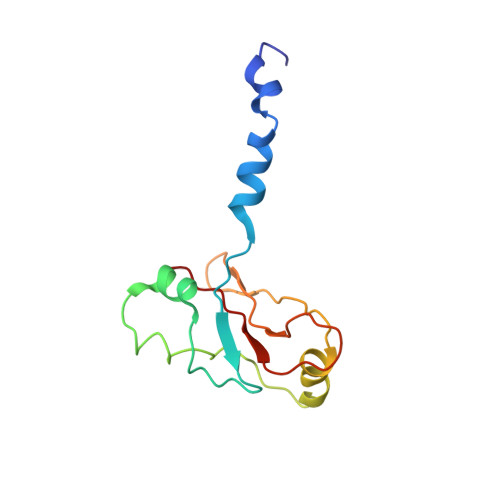
2W0N - PubMed Abstract:
The mechanistic understanding of how membrane-embedded sensor kinases recognize signals and regulate kinase activity is currently limited. Here we report structure-function relationships of the multidomain membrane sensor kinase DcuS using solid-state NMR, structural modeling and mutagenesis. Experimental data of an individual cytoplasmic Per-Arnt-Sim (PAS) domain were compared to structural models generated in silico. These studies, together with previous NMR work on the periplasmic PAS domain, enabled structural investigations of a membrane-embedded 40-kDa construct by solid-state NMR, comprising both PAS segments and the membrane domain. Structural alterations are largely limited to protein regions close to the transmembrane segment. Data from isolated and multidomain constructs favor a disordered N-terminal helix in the cytoplasmic domain. Mutations of residues in this region strongly influence function, suggesting that protein flexibility is related to signal transduction toward the kinase domain and regulation of kinase activity.
Organizational Affiliation:
Max-Planck-Institute for Biophysical Chemistry, Department of NMR-Based Structural Biology, Am Fassberg 11, 37077 Göttingen, Germany.