2W0C

X-ray structure of the entire lipid-containing bacteriophage PM2


Experimental Data Snapshot

  • Method: X-RAY DIFFRACTION
  • Resolution: 7.00 Å
  • R-Value Work: 0.419 
  • R-Value Observed: 0.419 

wwPDB Validation   3D Report Full Report


This is version 1.3 of the entry. See complete history


Literature

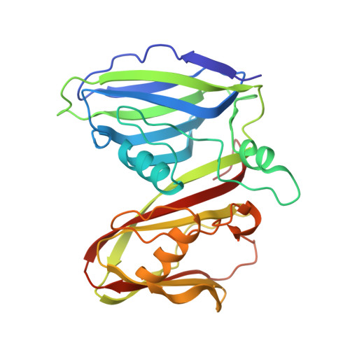
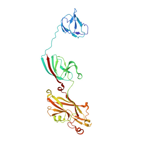
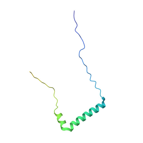
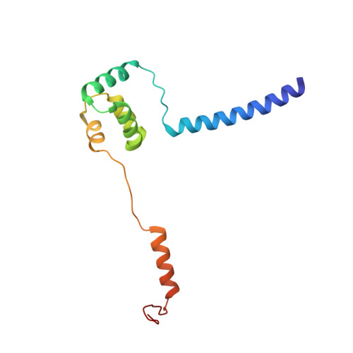
Insights Into Virus Evolution and Membrane Biogenesis from the Structure of the Marine Lipid-Containing Bacteriophage Pm2

Abrescia, N.G.A.Grimes, J.M.Kivela, H.M.Assenberg, R.Sutton, G.C.Butcher, S.J.Bamford, J.K.H.Bamford, D.H.Stuart, D.I.

(2008) Mol Cell 31: 749

  • DOI: https://doi.org/10.1016/j.molcel.2008.06.026
  • Primary Citation of Related Structures:  
    2VVD, 2VVE, 2VVF, 2W0C

  • PubMed Abstract: 

    Recent, primarily structural observations indicate that related viruses, harboring no sequence similarity, infect hosts of different domains of life. One such clade of viruses, defined by common capsid architecture and coat protein fold, is the so-called PRD1-adenovirus lineage. Here we report the structure of the marine lipid-containing bacteriophage PM2 determined by crystallographic analyses of the entire approximately 45 MDa virion and of the outer coat proteins P1 and P2, revealing PM2 to be a primeval member of the PRD1-adenovirus lineage with an icosahedral shell and canonical double beta barrel major coat protein. The view of the lipid bilayer, richly decorated with membrane proteins, constitutes a rare visualization of an in vivo membrane. The viral membrane proteins P3 and P6 are organized into a lattice, suggesting a possible assembly pathway to produce the mature virus.


  • Organizational Affiliation

    Division of Structural Biology and the Oxford Protein Production Facility, The Wellcome Trust Centre for Human Genetics, University of Oxford, Roosevelt Drive, Headington, Oxford OX3 7BN, UK.


Macromolecules
Find similar proteins by:  (by identity cutoff)  |  3D Structure
Entity ID: 1
MoleculeChains Sequence LengthOrganismDetailsImage
MAJOR CAPSID PROTEIN P2
A, B, C, D, E
A, B, C, D, E, F, G, H, I, J
269Corticovirus PM2Mutation(s): 0 
UniProt
Find proteins for P15794 (Pseudoalteromonas phage PM2)
Explore P15794 
Go to UniProtKB:  P15794
Entity Groups  
Sequence Clusters30% Identity50% Identity70% Identity90% Identity95% Identity100% Identity
UniProt GroupP15794
Sequence Annotations
Expand
  • Reference Sequence
Find similar proteins by:  (by identity cutoff)  |  3D Structure
Entity ID: 2
MoleculeChains Sequence LengthOrganismDetailsImage
PROTEIN 2K [auth L]335Corticovirus PM2Mutation(s): 0 
UniProt
Find proteins for Q9XJR3 (Pseudoalteromonas phage PM2)
Explore Q9XJR3 
Go to UniProtKB:  Q9XJR3
Entity Groups  
Sequence Clusters30% Identity50% Identity70% Identity90% Identity95% Identity100% Identity
UniProt GroupQ9XJR3
Sequence Annotations
Expand
  • Reference Sequence
Find similar proteins by:  (by identity cutoff)  |  3D Structure
Entity ID: 3
MoleculeChains Sequence LengthOrganismDetailsImage
PROTEIN P3L [auth P],
M [auth Q],
N [auth R],
O [auth S]
104Corticovirus PM2Mutation(s): 0 
UniProt
Find proteins for Q9XJR6 (Pseudoalteromonas phage PM2)
Explore Q9XJR6 
Go to UniProtKB:  Q9XJR6
Entity Groups  
Sequence Clusters30% Identity50% Identity70% Identity90% Identity95% Identity100% Identity
UniProt GroupQ9XJR6
Sequence Annotations
Expand
  • Reference Sequence
Find similar proteins by:  (by identity cutoff)  |  3D Structure
Entity ID: 4
MoleculeChains Sequence LengthOrganismDetailsImage
PROTEIN P6P [auth T]127Corticovirus PM2Mutation(s): 0 
UniProt
Find proteins for Q9XJR1 (Pseudoalteromonas phage PM2)
Explore Q9XJR1 
Go to UniProtKB:  Q9XJR1
Entity Groups  
Sequence Clusters30% Identity50% Identity70% Identity90% Identity95% Identity100% Identity
UniProt GroupQ9XJR1
Sequence Annotations
Expand
  • Reference Sequence
Small Molecules
Ligands 1 Unique
IDChains Name / Formula / InChI Key2D Diagram3D Interactions
CA
Query on CA

Download Ideal Coordinates CCD File 
AA [auth L]
Q [auth A]
R [auth B]
S [auth C]
T [auth D]
AA [auth L],
Q [auth A],
R [auth B],
S [auth C],
T [auth D],
U [auth E],
V [auth F],
W [auth G],
X [auth H],
Y [auth I],
Z [auth J]
CALCIUM ION
Ca
BHPQYMZQTOCNFJ-UHFFFAOYSA-N
Experimental Data & Validation

Experimental Data

  • Method: X-RAY DIFFRACTION
  • Resolution: 7.00 Å
  • R-Value Work: 0.419 
  • R-Value Observed: 0.419 
  • Space Group: C 1 2 1
Unit Cell:
Length ( Å )Angle ( ˚ )
a = 946.9α = 90
b = 677.6β = 102.9
c = 1067.6γ = 90
Software Package:
Software NamePurpose
X-PLORrefinement
DENZOdata reduction
SCALEPACKdata scaling
X-PLORphasing

Structure Validation

View Full Validation Report


Entry History 

Deposition Data

Revision History  (Full details and data files)

  • Version 1.0: 2008-09-23
    Type: Initial release
  • Version 1.1: 2011-07-07
    Changes: Version format compliance
  • Version 1.2: 2011-07-13
    Changes: Version format compliance
  • Version 1.3: 2017-10-18
    Changes: Data collection