2VZY

Crystal structure of Rv0802c from Mycobacterium tuberculosis in an unliganded form.


Experimental Data Snapshot

  • Method: X-RAY DIFFRACTION
  • Resolution: 2.00 Å
  • R-Value Free: 0.205 
  • R-Value Work: 0.164 
  • R-Value Observed: 0.166 

wwPDB Validation   3D Report Full Report


This is version 1.1 of the entry. See complete history


Literature

Rv0802C from Mycobacterium Tuberculosis: The First Structure of a Succinyltransferase with the Gnat Fold.

Vetting, M.W.Errey, J.C.Blanchard, J.S.

(2008) Acta Crystallogr Sect F Struct Biol Cryst Commun 64: 978

  • DOI: https://doi.org/10.1107/S1744309108031679
  • Primary Citation of Related Structures:  
    2VZY, 2VZZ

  • PubMed Abstract: 

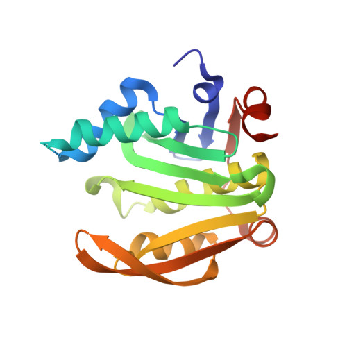
    Gene rv0802c from Mycobacterium tuberculosis encodes a 218-amino-acid protein and is annotated as a hypothetical protein with homology to GCN5-related N-acetyltransferases. The structure of Rv0802c was determined in an unliganded form to 2.0 A resolution utilizing single-wavelength anomalous dispersion from a samarium soak that resulted in a single bound Sm(3+):citrate(2) complex. The structure confirms that Rv0802c exhibits the GCN5-related N-acetyltransferase fold and revealed a tetramer composed of a dimer of dimers with approximate 222 symmetry. In addition, a bound acetate ion indicated that Rv0802c may utilize a unique acyl donor for the family. The subsequent determination of the structure of Rv0802c in complex with succinyl-CoA to 2.3 A resolution suggests that Rv0802c is the first known GCN5-related N-acetyltransferase family member to utilize succinyl-CoA as a substrate.


  • Organizational Affiliation

    Department of Biochemistry, Albert Einstein College of Medicine, 1300 Morris Park Avenue, Bronx, NY 10461, USA. vetting@aecom.yu.edu


Macromolecules
Find similar proteins by:  (by identity cutoff)  |  3D Structure
Entity ID: 1
MoleculeChains Sequence LengthOrganismDetailsImage
RV0802C
A, B, C, D
218Mycobacterium tuberculosis H37RvMutation(s): 0 
UniProt
Find proteins for P9WQG7 (Mycobacterium tuberculosis (strain ATCC 25618 / H37Rv))
Explore P9WQG7 
Go to UniProtKB:  P9WQG7
Entity Groups  
Sequence Clusters30% Identity50% Identity70% Identity90% Identity95% Identity100% Identity
UniProt GroupP9WQG7
Sequence Annotations
Expand
  • Reference Sequence
Small Molecules
Ligands 5 Unique
IDChains Name / Formula / InChI Key2D Diagram3D Interactions
FLC
Query on FLC

Download Ideal Coordinates CCD File 
Q [auth D],
R [auth D]
CITRATE ANION
C6 H5 O7
KRKNYBCHXYNGOX-UHFFFAOYSA-K
SM
Query on SM

Download Ideal Coordinates CCD File 
T [auth D]SAMARIUM (III) ION
Sm
DOSGOCSVHPUUIA-UHFFFAOYSA-N
SO4
Query on SO4

Download Ideal Coordinates CCD File 
F [auth A],
I [auth B],
L [auth C],
P [auth D]
SULFATE ION
O4 S
QAOWNCQODCNURD-UHFFFAOYSA-L
ETX
Query on ETX

Download Ideal Coordinates CCD File 
G [auth A],
J [auth B],
M [auth C],
N [auth C],
S [auth D]
2-ETHOXYETHANOL
C4 H10 O2
ZNQVEEAIQZEUHB-UHFFFAOYSA-N
ACT
Query on ACT

Download Ideal Coordinates CCD File 
E [auth A],
H [auth B],
K [auth C],
O [auth D]
ACETATE ION
C2 H3 O2
QTBSBXVTEAMEQO-UHFFFAOYSA-M
Experimental Data & Validation

Experimental Data

  • Method: X-RAY DIFFRACTION
  • Resolution: 2.00 Å
  • R-Value Free: 0.205 
  • R-Value Work: 0.164 
  • R-Value Observed: 0.166 
  • Space Group: P 21 21 21
Unit Cell:
Length ( Å )Angle ( ˚ )
a = 70.705α = 90
b = 111.458β = 90
c = 135.238γ = 90
Software Package:
Software NamePurpose
REFMACrefinement
DENZOdata reduction
SCALEPACKdata scaling
SOLVEphasing

Structure Validation

View Full Validation Report


Entry History 

Deposition Data

Revision History  (Full details and data files)

  • Version 1.0: 2009-04-07
    Type: Initial release
  • Version 1.1: 2011-07-13
    Changes: Advisory, Version format compliance