2VYR

Structure of human MDM4 N-terminal domain bound to a single domain antibody


Experimental Data Snapshot

  • Method: X-RAY DIFFRACTION
  • Resolution: 2.00 Å
  • R-Value Free: 0.248 
  • R-Value Work: 0.207 

wwPDB Validation   3D Report Full Report


This is version 1.1 of the entry. See complete history


Literature

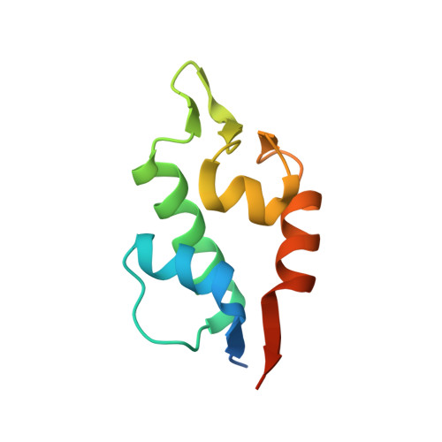
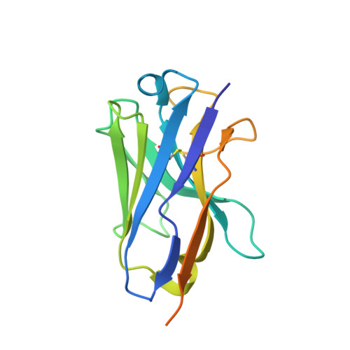
Structure of Human Mdm4 N-Terminal Domain Bound to a Single-Domain Antibody.

Yu, G.W.Vaysburd, M.Allen, M.D.Settanni, G.Fersht, A.R.

(2009) J Mol Biol 385: 1578

  • DOI: https://doi.org/10.1016/j.jmb.2008.11.043
  • Primary Citation of Related Structures:  
    2VYR

  • PubMed Abstract: 

    The N-terminal domain of MDM4 binds to the N-terminal transactivation domain of the tumor suppressor p53 and is an important negative regulator of its transactivation activity. As such, inhibition of the binding of MDM4 to p53 is a target for anticancer therapy. The protein has not been crystallized satisfactorily for structural studies without the addition of an N-terminal p53 peptide. We selected a single-domain antibody (VH9) that bound to the human domain with a dissociation constant of 44 nM. We solved the structure of the complex at 2.0-A resolution. The asymmetric unit contained eight molecules of VH9 and four molecules of MDM4. A molecule of VH9 was located in each transactivation domain binding site, and the four non-MDM4-bound VH9 domains provided additional crystal contacts. There are differences between the structures of human MDM4 domain bound to VH9 and those of human and zebra fish MDM4 bound to a p53 peptide. Molecular dynamics simulations showed that the binding pocket in the three MDM4 structures converged to a common conformation after removal of the ligands, indicating that the differences are due to induced fit. The largest conformational changes were for the MDM4 molecules bound to p53. The simulated and observed structures should aid rational drug design. The use of single-domain antibodies to aid crystallization by creating a molecular scaffold may have a wider range of applications.


  • Organizational Affiliation

    Centre for Protein Engineering, Medical Research Council, Cambridge, UK.


Macromolecules
Find similar proteins by:  (by identity cutoff)  |  3D Structure
Entity ID: 1
MoleculeChains Sequence LengthOrganismDetailsImage
MDM4 PROTEIN
A, B, C, D
101Homo sapiensMutation(s): 0 
UniProt & NIH Common Fund Data Resources
Find proteins for O15151 (Homo sapiens)
Explore O15151 
Go to UniProtKB:  O15151
PHAROS:  O15151
GTEx:  ENSG00000198625 
Entity Groups  
Sequence Clusters30% Identity50% Identity70% Identity90% Identity95% Identity100% Identity
UniProt GroupO15151
Sequence Annotations
Expand
  • Reference Sequence
Find similar proteins by:  (by identity cutoff)  |  3D Structure
Entity ID: 2
MoleculeChains Sequence LengthOrganismDetailsImage
HUMAN SINGLE DOMAIN ANTIBODY
E, F, G, H, I
E, F, G, H, I, J, K, L
153Homo sapiensMutation(s): 0 
Entity Groups  
Sequence Clusters30% Identity50% Identity70% Identity90% Identity95% Identity100% Identity
Sequence Annotations
Expand
  • Reference Sequence
Experimental Data & Validation

Experimental Data

  • Method: X-RAY DIFFRACTION
  • Resolution: 2.00 Å
  • R-Value Free: 0.248 
  • R-Value Work: 0.207 
  • Space Group: P 1 21 1
Unit Cell:
Length ( Å )Angle ( ˚ )
a = 79.823α = 90
b = 115.2β = 104.86
c = 99.368γ = 90
Software Package:
Software NamePurpose
MOSFLMdata reduction
SCALAdata scaling
SHELXphasing
PHASERphasing
PHENIXrefinement

Structure Validation

View Full Validation Report


Entry History 

Deposition Data

Revision History  (Full details and data files)

  • Version 1.0: 2008-11-25
    Type: Initial release
  • Version 1.1: 2013-03-06
    Changes: Data collection, Database references, Derived calculations, Non-polymer description, Other, Source and taxonomy, Structure summary, Version format compliance