2VYN

Structure of E.Coli GAPDH Rat Sperm GAPDH heterotetramer


Experimental Data Snapshot

  • Method: X-RAY DIFFRACTION
  • Resolution: 2.20 Å
  • R-Value Free: 0.224 
  • R-Value Work: 0.157 
  • R-Value Observed: 0.160 

wwPDB Validation   3D Report Full Report


Ligand Structure Quality Assessment 


This is version 1.4 of the entry. See complete history


Literature

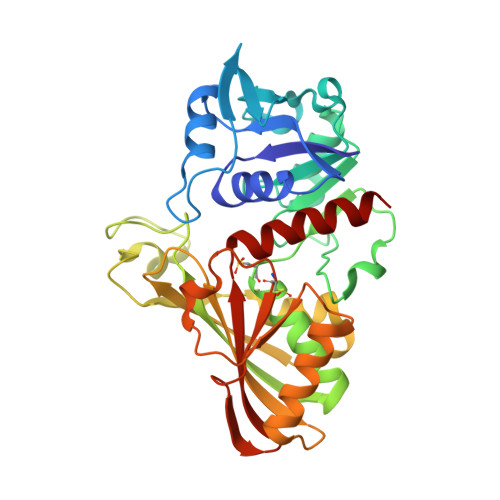
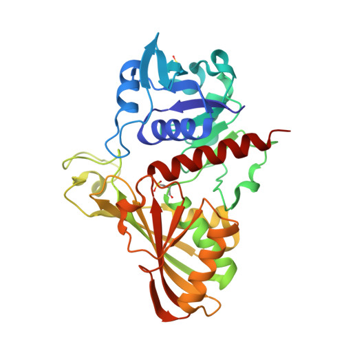
Structure of Insoluble Rat Sperm Glyceraldehyde-3-Phosphate Dehydrogenase (Gapdh) Via Heterotetramer Formation with Escherichia Coli Gapdh Reveals Target for Contraceptive Design.

Frayne, J.Taylor, A.Cameron, G.Hadfield, A.T.

(2009) J Biol Chem 284: 22703

  • DOI: https://doi.org/10.1074/jbc.M109.004648
  • Primary Citation of Related Structures:  
    2VYN, 2VYV

  • PubMed Abstract: 

    Sperm glyceraldehyde-3-phosphate dehydrogenase has been shown to be a successful target for a non-hormonal contraceptive approach, but the agents tested to date have had unacceptable side effects. Obtaining the structure of the sperm-specific isoform to allow rational inhibitor design has therefore been a goal for a number of years but has proved intractable because of the insoluble nature of both native and recombinant protein. We have obtained soluble recombinant sperm glyceraldehyde-3-phosphate dehydrogenase as a heterotetramer with the Escherichia coli glyceraldehyde-3-phosphate dehydrogenase in a ratio of 1:3 and have solved the structure of the heterotetramer which we believe represents a novel strategy for structure determination of an insoluble protein. A structure was also obtained where glyceraldehyde 3-phosphate binds in the P(s) pocket in the active site of the sperm enzyme subunit in the presence of NAD. Modeling and comparison of the structures of human somatic and sperm-specific glyceraldehyde-3-phosphate dehydrogenase revealed few differences at the active site and hence rebut the long presumed structural specificity of 3-chlorolactaldehyde for the sperm isoform. The contraceptive activity of alpha-chlorohydrin and its apparent specificity for the sperm isoform in vivo are likely to be due to differences in metabolism to 3-chlorolactaldehyde in spermatozoa and somatic cells. However, further detailed analysis of the sperm glyceraldehyde-3-phosphate dehydrogenase structure revealed sites in the enzyme that do show significant difference compared with published somatic glyceraldehyde-3-phosphate dehydrogenase structures that could be exploited by structure-based drug design to identify leads for novel male contraceptives.


  • Organizational Affiliation

    Department of Biochemistry, University of Bristol School of Medical Sciences, University Walk, Bristol BS8 1TD, United Kingdom. jan.frayne@bristol.ac.uk


Macromolecules
Find similar proteins by:  (by identity cutoff)  |  3D Structure
Entity ID: 1
MoleculeChains Sequence LengthOrganismDetailsImage
GLYCERALDEHYDE-3-PHOSPHATE DEHYDROGENASE
A, B, C
331Escherichia coli BL21(DE3)Mutation(s): 0 
EC: 1.2.1.12
UniProt
Find proteins for P0A9B2 (Escherichia coli (strain K12))
Explore P0A9B2 
Go to UniProtKB:  P0A9B2
Entity Groups  
Sequence Clusters30% Identity50% Identity70% Identity90% Identity95% Identity100% Identity
UniProt GroupP0A9B2
Sequence Annotations
Expand
  • Reference Sequence
Find similar proteins by:  (by identity cutoff)  |  3D Structure
Entity ID: 2
MoleculeChains Sequence LengthOrganismDetailsImage
GLYCERALDEHYDE-3-PHOSPHATE DEHYDROGENASE334Rattus norvegicusMutation(s): 0 
EC: 1.2.1.12
UniProt
Find proteins for Q9ESV6 (Rattus norvegicus)
Explore Q9ESV6 
Go to UniProtKB:  Q9ESV6
Entity Groups  
Sequence Clusters30% Identity50% Identity70% Identity90% Identity95% Identity100% Identity
UniProt GroupQ9ESV6
Sequence Annotations
Expand
  • Reference Sequence
Small Molecules
Ligands 2 Unique
IDChains Name / Formula / InChI Key2D Diagram3D Interactions
NAD
Query on NAD

Download Ideal Coordinates CCD File 
H [auth A],
M [auth B],
P [auth C],
V [auth D]
NICOTINAMIDE-ADENINE-DINUCLEOTIDE
C21 H27 N7 O14 P2
BAWFJGJZGIEFAR-NNYOXOHSSA-N
FMT
Query on FMT

Download Ideal Coordinates CCD File 
E [auth A]
F [auth A]
G [auth A]
I [auth B]
J [auth B]
E [auth A],
F [auth A],
G [auth A],
I [auth B],
J [auth B],
K [auth B],
L [auth B],
N [auth C],
O [auth C],
Q [auth D],
R [auth D],
S [auth D],
T [auth D],
U [auth D]
FORMIC ACID
C H2 O2
BDAGIHXWWSANSR-UHFFFAOYSA-N
Modified Residues  2 Unique
IDChains TypeFormula2D DiagramParent
CSD
Query on CSD
A, B, C
L-PEPTIDE LINKINGC3 H7 N O4 SCYS
CSX
Query on CSX
D
L-PEPTIDE LINKINGC3 H7 N O3 SCYS
Experimental Data & Validation

Experimental Data

  • Method: X-RAY DIFFRACTION
  • Resolution: 2.20 Å
  • R-Value Free: 0.224 
  • R-Value Work: 0.157 
  • R-Value Observed: 0.160 
  • Space Group: P 21 21 21
Unit Cell:
Length ( Å )Angle ( ˚ )
a = 68.445α = 90
b = 103.562β = 90
c = 177.832γ = 90
Software Package:
Software NamePurpose
REFMACrefinement
HKL-2000data reduction
HKL-2000data scaling
PHASERphasing

Structure Validation

View Full Validation Report


Ligand Structure Quality Assessment 


Entry History 

Deposition Data

Revision History  (Full details and data files)

  • Version 1.0: 2008-08-05
    Type: Initial release
  • Version 1.1: 2011-05-07
    Changes: Version format compliance
  • Version 1.2: 2011-07-13
    Changes: Version format compliance
  • Version 1.3: 2018-10-31
    Changes: Data collection, Source and taxonomy
  • Version 1.4: 2023-12-13
    Changes: Data collection, Database references, Derived calculations, Other, Refinement description