2VYE

Crystal Structure of the DnaC-ssDNA complex


Experimental Data Snapshot

  • Method: X-RAY DIFFRACTION
  • Resolution: 4.10 Å
  • R-Value Free: 0.374 
  • R-Value Work: 0.302 
  • R-Value Observed: 0.302 

wwPDB Validation   3D Report Full Report


This is version 1.3 of the entry. See complete history


Literature

The Crystal Structure of a Replicative Hexameric Helicase Dnac and its Complex with Single-Stranded DNA.

Lo, Y.H.Tsai, K.L.Sun, Y.J.Chen, W.T.Huang, C.Y.Hsiao, C.D.

(2009) Nucleic Acids Res 37: 804

  • DOI: https://doi.org/10.1093/nar/gkn999
  • Primary Citation of Related Structures:  
    2VYE, 2VYF

  • PubMed Abstract: 

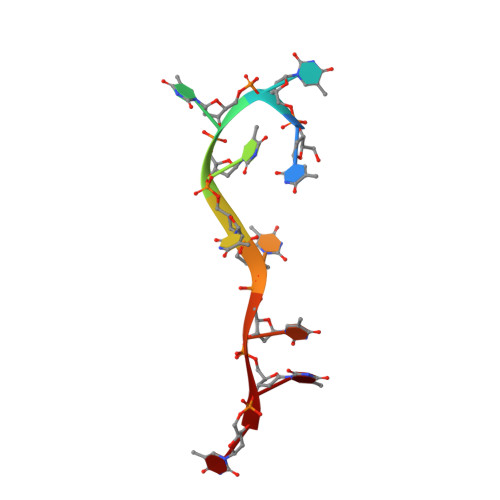
    DNA helicases are motor proteins that play essential roles in DNA replication, repair and recombination. In the replicative hexameric helicase, the fundamental reaction is the unwinding of duplex DNA; however, our understanding of this function remains vague due to insufficient structural information. Here, we report two crystal structures of the DnaB-family replicative helicase from Geobacillus kaustophilus HTA426 (GkDnaC) in the apo-form and bound to single-stranded DNA (ssDNA). The GkDnaC-ssDNA complex structure reveals that three symmetrical basic grooves on the interior surface of the hexamer individually encircle ssDNA. The ssDNA-binding pockets in this structure are directed toward the N-terminal domain collar of the hexameric ring, thus orienting the ssDNA toward the DnaG primase to facilitate the synthesis of short RNA primers. These findings provide insight into the mechanism of ssDNA binding and provide a working model to establish a novel mechanism for DNA translocation at the replication fork.


  • Organizational Affiliation

    Institute of Molecular Biology, Academia Sinica, Taipei, 115, Taiwan.


Macromolecules

Find similar proteins by:  (by identity cutoff)  |  3D Structure
Entity ID: 1
MoleculeChains Sequence LengthOrganismDetailsImage
REPLICATIVE DNA HELICASE
A, B
454Geobacillus kaustophilus HTA426Mutation(s): 0 
EC: 3.6.1
UniProt
Find proteins for Q5KU75 (Geobacillus kaustophilus (strain HTA426))
Explore Q5KU75 
Go to UniProtKB:  Q5KU75
Entity Groups  
Sequence Clusters30% Identity50% Identity70% Identity90% Identity95% Identity100% Identity
UniProt GroupQ5KU75
Sequence Annotations
Expand
  • Reference Sequence

Find similar nucleic acids by:  Sequence   |   3D Structure  

Entity ID: 2
MoleculeChains LengthOrganismImage
5'-D(*TP*TP*TP*TP*TP*TP*TP*TP*TP)-3'9Escherichia coli
Sequence Annotations
Expand
  • Reference Sequence
Experimental Data & Validation

Experimental Data

  • Method: X-RAY DIFFRACTION
  • Resolution: 4.10 Å
  • R-Value Free: 0.374 
  • R-Value Work: 0.302 
  • R-Value Observed: 0.302 
  • Space Group: P 63
Unit Cell:
Length ( Å )Angle ( ˚ )
a = 180.83α = 90
b = 180.83β = 90
c = 104.112γ = 120
Software Package:
Software NamePurpose
CNSrefinement
SCALEPACKdata scaling
MOLREPphasing

Structure Validation

View Full Validation Report


Entry History 

Deposition Data

Revision History  (Full details and data files)

  • Version 1.0: 2008-12-30
    Type: Initial release
  • Version 1.1: 2012-05-16
    Changes: Advisory, Database references, Derived calculations, Other, Refinement description, Structure summary, Version format compliance
  • Version 1.2: 2018-01-24
    Changes: Source and taxonomy
  • Version 1.3: 2023-12-13
    Changes: Data collection, Database references, Other, Refinement description