Crystal Structure of the Botulinum Neurotoxin Type G Binding Domain: Insight Into Cell Surface Binding.
Stenmark, P., Dong, M., Dupuy, J., Chapman, E.R., Stevens, R.C.(2010) J Mol Biol 397: 1287
- PubMed: 20219474
- DOI: https://doi.org/10.1016/j.jmb.2010.02.041
- Primary Citation of Related Structures:
2VXR - PubMed Abstract:
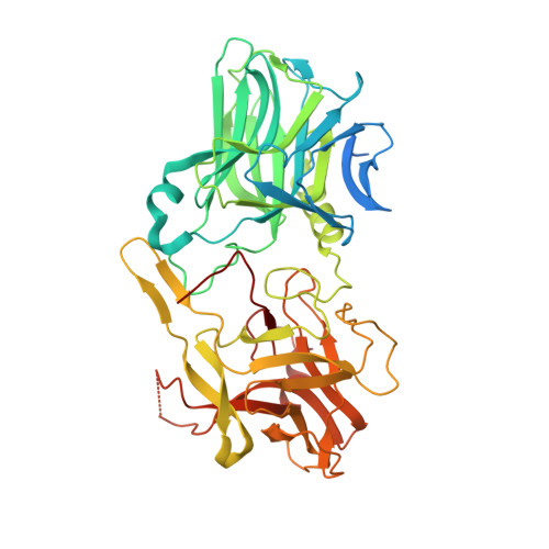
Botulinum neurotoxins (BoNTs) typically bind the neuronal cell surface via dual interactions with both protein receptors and gangliosides. We present here the 1.9-A X-ray structure of the BoNT serotype G (BoNT/G) receptor binding domain (residues 868-1297) and a detailed view of protein receptor and ganglioside binding regions. The ganglioside binding motif (SxWY) has a conserved structure compared to the corresponding regions in BoNT serotype A and BoNT serotype B (BoNT/B), but several features of interactions with the hydrophilic face of the ganglioside are absent at the opposite side of the motif in the BoNT/G ganglioside binding cleft. This may significantly reduce the affinity between BoNT/G and gangliosides. BoNT/G and BoNT/B share the protein receptor synaptotagmin (Syt) I/II. The Syt binding site has a conserved hydrophobic plateau located centrally in the proposed protein receptor binding interface (Tyr1189, Phe1202, Ala1204, Pro1205, and Phe1212). Interestingly, only 5 of 14 residues that are important for binding between Syt-II and BoNT/B are conserved in BoNT/G, suggesting that the means by which BoNT/G and BoNT/B bind Syt diverges more than previously appreciated. Indeed, substitution of Syt-II Phe47 and Phe55 with alanine residues had little effect on the binding of BoNT/G, but strongly reduced the binding of BoNT/B. Furthermore, an extended solvent-exposed hydrophobic loop, located between the Syt binding site and the ganglioside binding cleft, may serve as a third membrane association and binding element to contribute to high-affinity binding to the neuronal membrane. While BoNT/G and BoNT/B are homologous to each other and both utilize Syt-I/Syt-II as their protein receptor, the precise means by which these two toxin serotypes bind to Syt appears surprisingly divergent.
Organizational Affiliation:
Department of Molecular Biology, The Scripps Research Institute, 10550 North Torrey Pines Road, La Jolla, CA 92037, USA.