Changes in Quaternary Structure in the Signaling Mechanisms of Pas Domains.
Ayers, R.A., Moffat, K.(2008) Biochemistry 47: 12078
- PubMed: 18942854
- DOI: https://doi.org/10.1021/bi801254c
- Primary Citation of Related Structures:
2VV6, 2VV7, 2VV8 - PubMed Abstract:
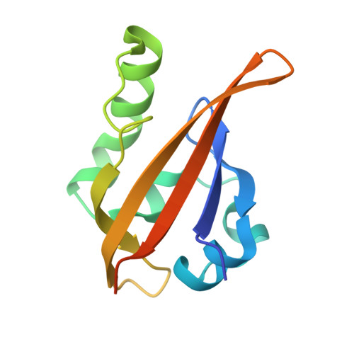
FixL from Bradyrhizobium japonicum is a PAS sensor protein in which two PAS domains covalently linked to a histidine kinase domain are responsible for regulating nitrogen fixation in an oxygen-dependent manner. The more C-terminal PAS domain, denoted bjFixLH, contains a heme cofactor that binds diatomic molecules such as carbon monoxide and oxygen and regulates the activity of the FixL histidine kinase as part of a two-component signaling system. We present the structures of ferric, deoxy, and carbon monoxide-bound bjFixLH in a new space group ( P1) and at resolutions (1.5-1.8 A) higher than the resolutions of those previously obtained. Interestingly, bjFixLH can form two different dimers (in P1 and R32 crystal forms) in the same crystallization solution, where the monomers in one dimer are rotated approximately 175 degrees relative to the second. This suggests that PAS monomers are plastic and that two quite distinct quaternary structures are closely similar in free energy. We use screw rotation analysis to carry out a quantitative pairwise comparison of PAS quaternary structures, which identifies five different relative orientations adopted by isolated PAS monomers. We conclude that PAS monomer arrangement is context-dependent and could differ depending on whether the PAS domains are isolated or are part of a full-length protein. Structurally homologous residues comprise a conserved dimer interface. Using network analysis, we find that the architecture of the PAS dimer interface is continuous rather than modular; the network of residues comprising the interface is strongly connected. A continuous dimer interface is consistent with the low dimer-monomer dissociation equilibrium constant. Finally, we quantitate quaternary structural changes induced by carbon monoxide binding to a bjFixLH dimer, in which monomers rotate by up to approximately 2 degrees relative to each other. We relate these changes to those in other dimeric PAS domains and discuss the role of quaternary structural changes in the signaling mechanisms of PAS sensor proteins.
Organizational Affiliation:
Department of Biochemistry and Molecular Biology, The University of Chicago, 929 East 57th Street, Chicago, Illinois 60637, USA.