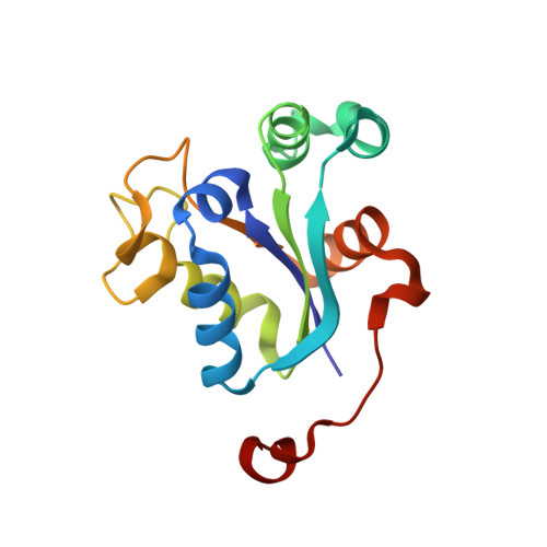
Crystal Structure of the Bacillus Anthracis Nucleoside Diphosphate Kinase and its Characterization Reveals an Enzyme Adapted to Perform Under Stress Conditions.
Misra, G., Aggarwal, A., Dube, D., Zaman, M.S., Singh, Y., Ramachandran, R.(2009) Proteins 76: 496
- PubMed: 19241473
- DOI: https://doi.org/10.1002/prot.22364
- Primary Citation of Related Structures:
2VU5 - PubMed Abstract:
Nucleoside diphosphate kinases (Ndks) play an important role in a plethora of regulatory and metabolic functions. Inhibition of the B. anthracis Ndk mRNA results in the formation of nonviable aberrant spores. We report the characterization and crystal structure of the enzyme from B. anthracis nucleoside diphosphate kinase (BaNdk), the first from sporulating bacteria. The enzyme, although from a mesophilic source, is active at extremes of pH (3.5-10.5), temperature (10-95 degrees C) and ionic strength (0.25-4.0M NaCl). It exists as a hexamer that is composed of two SDS-stable trimers interacting in a back-to-back association; mutational analysis confirms that the enzyme is a histidine kinase. The high-resolution crystal structure reported here reveals an unanticipated change in the conformation of residues between 43 and 63 that also regulates substrate entry in other Ndks. A comparative structural analysis involving Ndks from seven mesophiles and three thermophiles has resulted in the delineation of the structure into relatively rigid and flexible regions. The analysis suggests that the larger number of intramolecular hydrogen bonds and to a lesser extent ionic interactions in BaNdk contributes to its high thermostability. Mutational analysis and Molecular Dynamics simulations were used to probe the role of a highly conserved Gly19 (present at the oligomeric interface in most of the Ndks). The results suggest that the mutation leads to a rigidification of those residues that facilitate substrate entry and consequently leads to a large reduction in the kinase activity. Overall, the enzyme characterization helps to understand its apparent adaptation to perform under stress conditions.
Organizational Affiliation:
Central Drug Research Institute, Lucknow, India.