2VT1

Crystal structure of the cytoplasmic domain of Spa40, the specificity switch for the Shigella flexneri Type III Secretion System


Experimental Data Snapshot

  • Method: X-RAY DIFFRACTION
  • Resolution: 2.00 Å
  • R-Value Free: 0.227 
  • R-Value Work: 0.178 
  • R-Value Observed: 0.180 

wwPDB Validation   3D Report Full Report


This is version 1.4 of the entry. See complete history


Literature

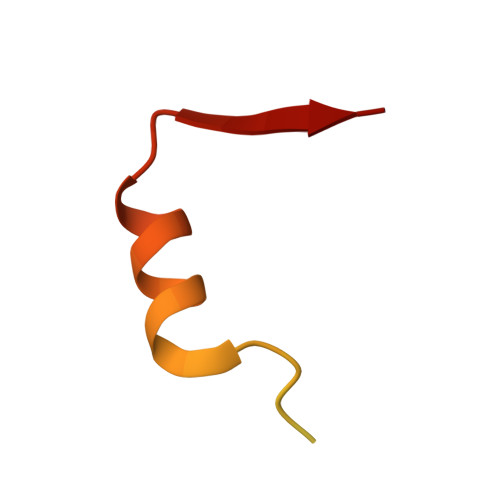
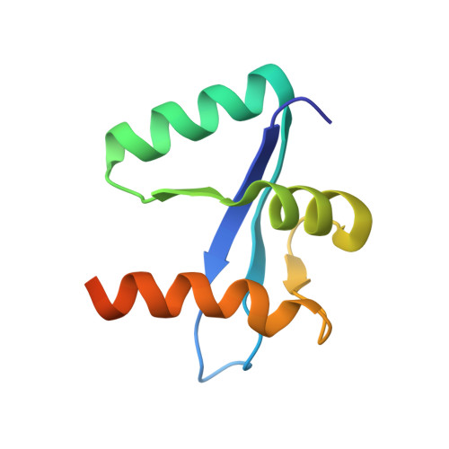
Crystal Structure of Spa40, the Specificity Switch for the Shigella Flexneri Type III Secretion System

Deane, J.E.Graham, S.C.Mitchell, E.P.Flot, D.Johnson, S.Lea, S.M.

(2008) Mol Microbiol 69: 267

  • DOI: https://doi.org/10.1111/j.1365-2958.2008.06293.x
  • Primary Citation of Related Structures:  
    2VT1

  • PubMed Abstract: 

    The pathogenic bacterium Shigella flexneri uses a type III secretion system to inject virulence factors from the bacterial cytosol directly into host cells. The machinery that identifies secretion substrates and controls the export of extracellular components and effector proteins consists of several inner-membrane and cytoplasmic proteins. One of the inner membrane components, Spa40, belongs to a family of proteins proposed to regulate the switching of substrate specificity of the export apparatus. We show that Spa40 is cleaved within the strictly conserved amino acid sequence NPTH and substitution of the proposed autocatalytic residue abolishes cleavage. Here we also report the crystal structure of the cytoplasmic complex Spa40(C) and compare it with the recent structures of the homologues from Escherichia coli and Salmonella typhimurium. These structures reveal the tight association of the cleaved fragments and show that the conserved NPTH sequence lies on a loop which, when cleaved, swings away from the catalytic N257 residue, resulting in different surface features in this region. This structural rearrangement suggests a mechanism by which non-cleaving forms of these proteins interfere with correct substrate switching of the apparatus.


  • Organizational Affiliation

    Sir William Dunn School of Pathology, South Parks Rd, University of Oxford, OX1 3RE, UK.


Macromolecules
Find similar proteins by:  (by identity cutoff)  |  3D Structure
Entity ID: 1
MoleculeChains Sequence LengthOrganismDetailsImage
SURFACE PRESENTATION OF ANTIGENS PROTEIN SPAS52Shigella flexneriMutation(s): 0 
UniProt
Find proteins for P0A1M8 (Shigella flexneri)
Explore P0A1M8 
Go to UniProtKB:  P0A1M8
Entity Groups  
Sequence Clusters30% Identity50% Identity70% Identity90% Identity95% Identity100% Identity
UniProt GroupP0A1M8
Sequence Annotations
Expand
  • Reference Sequence
Find similar proteins by:  (by identity cutoff)  |  3D Structure
Entity ID: 2
MoleculeChains Sequence LengthOrganismDetailsImage
SURFACE PRESENTATION OF ANTIGENS PROTEIN SPAS93Shigella flexneriMutation(s): 0 
UniProt
Find proteins for P0A1M8 (Shigella flexneri)
Explore P0A1M8 
Go to UniProtKB:  P0A1M8
Entity Groups  
Sequence Clusters30% Identity50% Identity70% Identity90% Identity95% Identity100% Identity
UniProt GroupP0A1M8
Sequence Annotations
Expand
  • Reference Sequence
Experimental Data & Validation

Experimental Data

  • Method: X-RAY DIFFRACTION
  • Resolution: 2.00 Å
  • R-Value Free: 0.227 
  • R-Value Work: 0.178 
  • R-Value Observed: 0.180 
  • Space Group: P 1
Unit Cell:
Length ( Å )Angle ( ˚ )
a = 25.035α = 102.52
b = 30.753β = 110.97
c = 32.099γ = 94.3
Software Package:
Software NamePurpose
REFMACrefinement
MOSFLMdata reduction
SCALAdata scaling
PHASERphasing

Structure Validation

View Full Validation Report


Entry History 

Deposition Data

Revision History  (Full details and data files)

  • Version 1.0: 2008-05-20
    Type: Initial release
  • Version 1.1: 2011-05-08
    Changes: Version format compliance
  • Version 1.2: 2011-07-13
    Changes: Version format compliance
  • Version 1.3: 2019-05-08
    Changes: Data collection, Experimental preparation, Other
  • Version 1.4: 2023-12-13
    Changes: Data collection, Database references, Other, Refinement description