2VSS

Wild-type Hydroxycinnamoyl-CoA hydratase lyase in complex with acetyl- CoA and vanillin


Experimental Data Snapshot

  • Method: X-RAY DIFFRACTION
  • Resolution: 2.22 Å
  • R-Value Free: 0.242 
  • R-Value Work: 0.183 
  • R-Value Observed: 0.186 

wwPDB Validation   3D Report Full Report


Ligand Structure Quality Assessment 


This is version 1.3 of the entry. See complete history


Literature

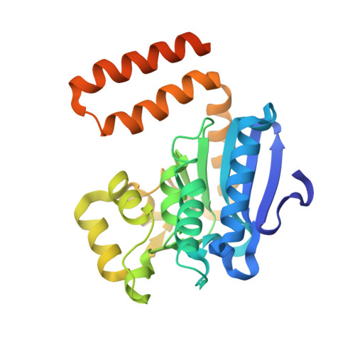
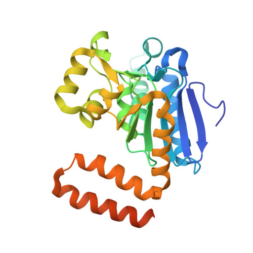
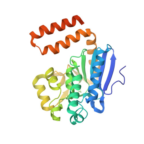
A Ternary Complex of Hydroxycinnamoyl-Coa Hydratase-Lyase (Hchl) with Acetyl-Coa and Vanillin Gives Insights Into Substrate Specificity and Mechanism.

Bennett, J.P.Bertin, L.M.Moulton, B.Fairlamb, I.J.S.Brzozowski, A.M.Walton, N.J.Grogan, G.

(2008) Biochem J 414: 281

  • DOI: https://doi.org/10.1042/BJ20080714
  • Primary Citation of Related Structures:  
    2VSS, 2VSU

  • PubMed Abstract: 

    HCHL (hydroxycinnamoyl-CoA hydratase-lyase) catalyses the biotransformation of feruloyl-CoA to acetyl-CoA and the important flavour-fragrance compound vanillin (4-hydroxy-3-methoxybenzaldehyde) and is exploited in whole-cell systems for the bioconversion of ferulic acid into natural equivalent vanillin. The reaction catalysed by HCHL has been thought to proceed by a two-step process involving first the hydration of the double bond of feruloyl-CoA and then the cleavage of the resultant beta-hydroxy thioester by retro-aldol reaction to yield the products. Kinetic analysis of active-site residues identified using the crystal structure of HCHL revealed that while Glu-143 was essential for activity, Ser-123 played no major role in catalysis. However, mutation of Tyr-239 to Phe greatly increased the K(M) for the substrate ferulic acid, fulfilling its anticipated role as a factor in substrate binding. Structures of WT (wild-type) HCHL and of the S123A mutant, each of which had been co-crystallized with feruloyl-CoA, reveal a subtle helix movement upon ligand binding, the consequence of which is to bring the phenolic hydroxyl of Tyr-239 into close proximity to Tyr-75 from a neighbouring subunit in order to bind the phenolic hydroxyl of the product vanillin, for which electron density was observed. The active-site residues of ligand-bound HCHL display a remarkable three-dimensional overlap with those of a structurally unrelated enzyme, vanillyl alcohol oxidase, that also recognizes p-hydroxylated aromatic substrates related to vanillin. The data both explain the observed substrate specificity of HCHL for p-hydroxylated cinnamate derivatives and illustrate a remarkable convergence of the molecular determinants of ligand recognition between the two otherwise unrelated enzymes.


  • Organizational Affiliation

    York Structural Biology Laboratory, Department of Chemistry, University of York, Heslington, York YO10 5YW, UK.


Macromolecules
Find similar proteins by:  (by identity cutoff)  |  3D Structure
Entity ID: 1
MoleculeChains Sequence LengthOrganismDetailsImage
P-HYDROXYCINNAMOYL COA HYDRATASE/LYASE
A, B, C, D
276Pseudomonas fluorescensMutation(s): 0 
EC: 4.2.1.101
UniProt
Find proteins for O69762 (Pseudomonas fluorescens)
Explore O69762 
Go to UniProtKB:  O69762
Entity Groups  
Sequence Clusters30% Identity50% Identity70% Identity90% Identity95% Identity100% Identity
UniProt GroupO69762
Sequence Annotations
Expand
  • Reference Sequence
Find similar proteins by:  (by identity cutoff)  |  3D Structure
Entity ID: 2
MoleculeChains Sequence LengthOrganismDetailsImage
P-HYDROXYCINNAMOYL COA HYDRATASE/LYASE276Pseudomonas fluorescensMutation(s): 0 
EC: 4.2.1.101
UniProt
Find proteins for O69762 (Pseudomonas fluorescens)
Explore O69762 
Go to UniProtKB:  O69762
Entity Groups  
Sequence Clusters30% Identity50% Identity70% Identity90% Identity95% Identity100% Identity
UniProt GroupO69762
Sequence Annotations
Expand
  • Reference Sequence
Find similar proteins by:  (by identity cutoff)  |  3D Structure
Entity ID: 3
MoleculeChains Sequence LengthOrganismDetailsImage
P-HYDROXYCINNAMOYL COA HYDRATASE/LYASE276Pseudomonas fluorescensMutation(s): 0 
EC: 4.2.1.101
UniProt
Find proteins for O69762 (Pseudomonas fluorescens)
Explore O69762 
Go to UniProtKB:  O69762
Entity Groups  
Sequence Clusters30% Identity50% Identity70% Identity90% Identity95% Identity100% Identity
UniProt GroupO69762
Sequence Annotations
Expand
  • Reference Sequence
Experimental Data & Validation

Experimental Data

  • Method: X-RAY DIFFRACTION
  • Resolution: 2.22 Å
  • R-Value Free: 0.242 
  • R-Value Work: 0.183 
  • R-Value Observed: 0.186 
  • Space Group: P 21 21 21
Unit Cell:
Length ( Å )Angle ( ˚ )
a = 90.223α = 90
b = 130.595β = 90
c = 144.663γ = 90
Software Package:
Software NamePurpose
REFMACrefinement
HKL-2000data reduction
SCALEPACKdata scaling
MOLREPphasing

Structure Validation

View Full Validation Report


Ligand Structure Quality Assessment 


Entry History 

Deposition Data

Revision History  (Full details and data files)

  • Version 1.0: 2008-05-27
    Type: Initial release
  • Version 1.1: 2011-07-13
    Changes: Advisory, Version format compliance
  • Version 1.2: 2022-05-04
    Changes: Database references, Derived calculations, Other, Structure summary
  • Version 1.3: 2023-12-13
    Changes: Data collection, Refinement description