2VQ2

Crystal structure of PilW, widely conserved type IV pilus biogenesis factor


Experimental Data Snapshot

  • Method: X-RAY DIFFRACTION
  • Resolution: 1.54 Å
  • R-Value Free: 0.183 
  • R-Value Work: 0.165 
  • R-Value Observed: 0.166 

wwPDB Validation   3D Report Full Report


This is version 1.4 of the entry. See complete history


Literature

Structure of a widely conserved type IV pilus biogenesis factor that affects the stability of secretin multimers.

Trindade, M.B.Job, V.Contreras-Martel, C.Pelicic, V.Dessen, A.

(2008) J Mol Biol 378: 1031-1039

  • DOI: https://doi.org/10.1016/j.jmb.2008.03.028
  • Primary Citation of Related Structures:  
    2VQ2

  • PubMed Abstract: 

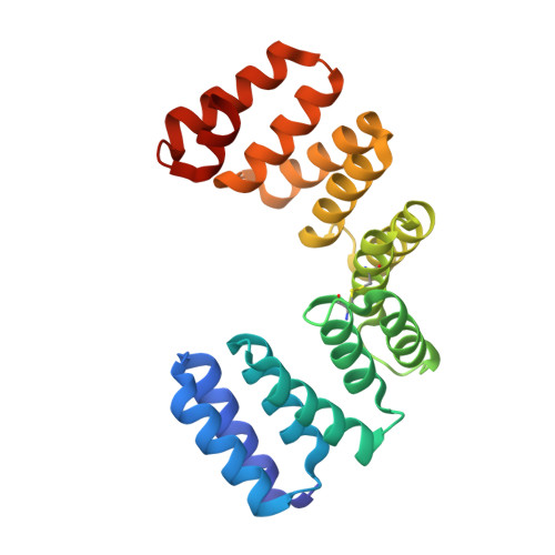
    Type IV pili (Tfp) are arguably the most widespread pili in bacteria, whose biogenesis requires a complex machinery composed of as many as 18 different proteins. This includes the conserved outer membrane-localized secretin, which forms a pore through which Tfp emerge on the bacterial surface. Although, in most model species studied, secretin oligomerization and functionality requires the action of partner lipoproteins, structural information regarding these molecules is limited. We report the high-resolution crystal structure of PilW, the partner lipoprotein of the type IV pilus secretin PilQ from Neisseria meningitidis, which defines a conserved class of Tfp biogenesis proteins involved in the formation and/or stability of secretin multimers in a wide variety of bacteria. The use of the PilW structure as a blueprint reveals an area of high-level sequence conservation in homologous proteins from different pathogens that could reflect a possible secretin-binding site. These results could be exploited for the development of new broad-spectrum antibacterials interfering with the biogenesis of a widespread virulence factor.


  • Organizational Affiliation

    Institut de Biologie Structurale Jean-Pierre Ebel, UMR 5075, 41 rue Jules Horowitz, F-38027 Grenoble, France.


Macromolecules
Find similar proteins by:  (by identity cutoff)  |  3D Structure
Entity ID: 1
MoleculeChains Sequence LengthOrganismDetailsImage
PUTATIVE FIMBRIAL BIOGENESIS AND TWITCHING MOTILITY PROTEIN225Neisseria meningitidisMutation(s): 0 
UniProt
Find proteins for Q9JZ41 (Neisseria meningitidis serogroup B (strain MC58))
Explore Q9JZ41 
Go to UniProtKB:  Q9JZ41
Entity Groups  
Sequence Clusters30% Identity50% Identity70% Identity90% Identity95% Identity100% Identity
UniProt GroupQ9JZ41
Sequence Annotations
Expand
  • Reference Sequence
Small Molecules
Ligands 5 Unique
IDChains Name / Formula / InChI Key2D Diagram3D Interactions
SO4
Query on SO4

Download Ideal Coordinates CCD File 
B [auth A]SULFATE ION
O4 S
QAOWNCQODCNURD-UHFFFAOYSA-L
GOL
Query on GOL

Download Ideal Coordinates CCD File 
C [auth A]GLYCEROL
C3 H8 O3
PEDCQBHIVMGVHV-UHFFFAOYSA-N
SO3
Query on SO3

Download Ideal Coordinates CCD File 
G [auth A]SULFITE ION
O3 S
LSNNMFCWUKXFEE-UHFFFAOYSA-L
CL
Query on CL

Download Ideal Coordinates CCD File 
D [auth A],
E [auth A],
F [auth A]
CHLORIDE ION
Cl
VEXZGXHMUGYJMC-UHFFFAOYSA-M
MG
Query on MG

Download Ideal Coordinates CCD File 
H [auth A]MAGNESIUM ION
Mg
JLVVSXFLKOJNIY-UHFFFAOYSA-N
Experimental Data & Validation

Experimental Data

  • Method: X-RAY DIFFRACTION
  • Resolution: 1.54 Å
  • R-Value Free: 0.183 
  • R-Value Work: 0.165 
  • R-Value Observed: 0.166 
  • Space Group: P 62 2 2
Unit Cell:
Length ( Å )Angle ( ˚ )
a = 87.446α = 90
b = 87.446β = 90
c = 117.102γ = 120
Software Package:
Software NamePurpose
REFMACrefinement
XDSdata reduction
XSCALEdata scaling
SHARPphasing

Structure Validation

View Full Validation Report


Entry History 

Deposition Data

Revision History  (Full details and data files)

  • Version 1.0: 2008-05-27
    Type: Initial release
  • Version 1.1: 2011-05-08
    Changes: Version format compliance
  • Version 1.2: 2011-07-13
    Changes: Version format compliance
  • Version 1.3: 2018-02-28
    Changes: Database references, Source and taxonomy
  • Version 1.4: 2019-05-15
    Changes: Data collection, Experimental preparation