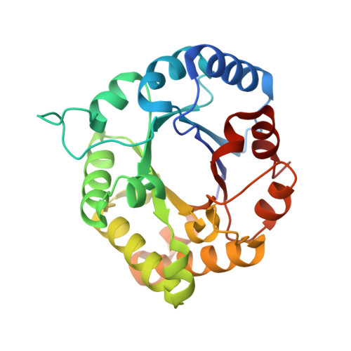
Structural Basis of Human Triosephosphate Isomerase Deficiency: Mutation E104D is Related to Alterations of a Conserved Water Network at the Dimer Interface.
Rodriguez-Almazan, C., Arreola-Alemon, R., Rodriguez-Larrea, D., Aguirre-Lopez, B., De Gomez-Puyou, M.T., Perez-Montfort, R., Costas, M., Gomez-Puyou, A., Torres-Larios, A.(2008) J Biol Chem 283: 23254
- PubMed: 18562316
- DOI: https://doi.org/10.1074/jbc.M802145200
- Primary Citation of Related Structures:
2JK2, 2VOM - PubMed Abstract:
Human triosephosphate isomerase deficiency is a rare autosomal disease that causes premature death of homozygous individuals. The most frequent mutation that leads to this illness is in position 104, which involves a conservative change of a Glu for Asp. Despite the extensive work that has been carried out on the E104D mutant enzyme in hemolysates and whole cells, the molecular basis of this disease is poorly understood. Here, we show that the purified, recombinant mutant enzyme E104D, while exhibiting normal catalytic activity, shows impairments in the formation of active dimers and low thermostability and monomerizes under conditions in which the wild type retains its dimeric form. The crystal structure of the E104D mutant at 1.85 A resolution showed that its global structure was similar to that of the wild type; however, residue 104 is part of a conserved cluster of 10 residues, five from each subunit. An analysis of the available high resolution structures of TIM dimers revealed that this cluster forms a cavity that possesses an elaborate conserved network of buried water molecules that bridge the two subunits. In the E104D mutant, a disruption of contacts of the amino acid side chains in the conserved cluster leads to a perturbation of the water network in which the water-protein and water-water interactions that join the two monomers are significantly weakened and diminished. Thus, the disruption of this solvent system would stand as the underlying cause of the deficiency.
Organizational Affiliation:
Departamento de Bioquímica, Instituto de Fisiología Celular, Universidad Nacional Autónoma de México, Ciudad Universitaria, Apartado Postal 70-243, Mexico City 04510, México.