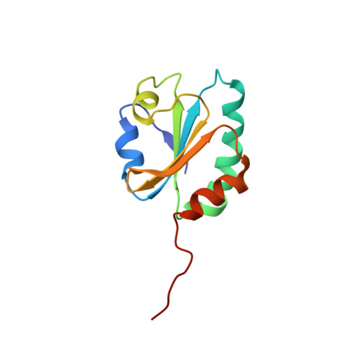
Thioredoxin A active-site mutants form mixed disulfide dimers that resemble enzyme-substrate reaction intermediates.
Kouwen, T.R., Andrell, J., Schrijver, R., Dubois, J.Y., Maher, M.J., Iwata, S., Carpenter, E.P., van Dijl, J.M.(2008) J Mol Biol 379: 520-534
- PubMed: 18455736
- DOI: https://doi.org/10.1016/j.jmb.2008.03.077
- Primary Citation of Related Structures:
2VOC - PubMed Abstract:
Thioredoxin functions in nearly all organisms as the major thiol-disulfide oxidoreductase within the cytosol. Its prime purpose is to maintain cysteine-containing proteins in the reduced state by converting intramolecular disulfide bonds into dithiols in a disulfide exchange reaction. Thioredoxin has been reported to contribute to a wide variety of physiological functions by interacting with specific sets of substrates in different cell types. To investigate the function of the essential thioredoxin A (TrxA) in the low-GC Gram-positive bacterium Bacillus subtilis, we purified wild-type TrxA and three mutant TrxA proteins that lack either one or both of the two cysteine residues in the CxxC active site. The pure proteins were used for substrate-binding studies known as "mixed disulfide fishing" in which covalent disulfide-bonded reaction intermediates can be visualized. An unprecedented finding is that both active-site cysteine residues can form mixed disulfides with substrate proteins when the other active-site cysteine is absent, but only the N-terminal active-site cysteine forms stable interactions. A second novelty is that both single-cysteine mutant TrxA proteins form stable homodimers due to thiol oxidation of the remaining active-site cysteine residue. To investigate whether these dimers resemble mixed enzyme-substrate disulfides, the structure of the most abundant dimer, C32S, was characterized by X-ray crystallography. This yielded a high-resolution (1.5A) X-ray crystallographic structure of a thioredoxin homodimer from a low-GC Gram-positive bacterium. The C32S TrxA dimer can be regarded as a mixed disulfide reaction intermediate of thioredoxin, which reveals the diversity of thioredoxin/substrate-binding modes.
Organizational Affiliation:
Department of Medical Microbiology, University Medical Center Groningen and University of Groningen, Hanzeplein 1, PO Box 30001, 9700 RB Groningen, The Netherlands.