2VN2

Crystal structure of the N-terminal domain of DnaD protein from Geobacillus kaustophilus HTA426


Experimental Data Snapshot

  • Method: X-RAY DIFFRACTION
  • Resolution: 2.30 Å
  • R-Value Free: 0.223 
  • R-Value Work: 0.217 
  • R-Value Observed: 0.217 

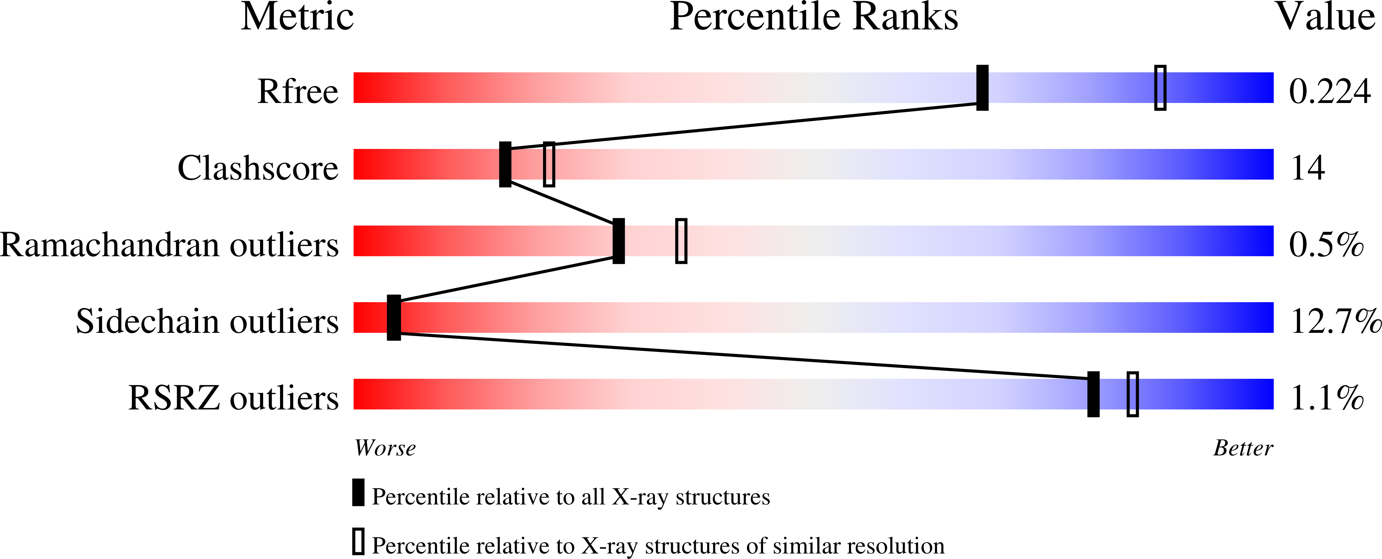
wwPDB Validation   3D Report Full Report


This is version 1.1 of the entry. See complete history


Literature

Crystal Structure of the N-Terminal Domain of Geobacillus Kaustophilus Hta426 Dnad Protein.

Huang, C.-Y.Chang, Y.-W.Chen, W.-T.

(2008) Biochem Biophys Res Commun 375: 220

  • DOI: https://doi.org/10.1016/j.bbrc.2008.07.160
  • Primary Citation of Related Structures:  
    2VN2

  • PubMed Abstract: 

    The DnaD is one of the primosomal proteins that are required for initiation and re-initiation of chromosomal DNA replication in Gram-positive bacteria. The DnaD protein is composed of two major structural domains: an N-terminal oligomerization domain and a C-terminal ssDNA binding domain. Here, we report the crystal structure of the N-terminal domain (aa 1-128) of DnaD (DnaDn) of Geobacillus kaustophilus HTA426 at 2.3A resolution. The structure of DnaDn reveals an extended winged-helix fold, a typical double-stranded DNA binding motif as winged-helix proteins. DnaDn formed tetramers in the crystalline state, but the results of gel filtration chromatography further indicated that this domain of DnaD was a stable dimer in solution. The structural analysis of DnaDn may suggest the binding sites for DNA and DnaB, and an assembly mechanism for Gram-positive bacterial DNA replication primosome.


  • Organizational Affiliation

    Department of Biomedical Sciences, Chung Shan Medical University, No. 110, Sec. 1, Chien-Kuo N. Road, Taichung 402, Taiwan. cyhuang@csmu.edu.tw


Macromolecules
Find similar proteins by:  (by identity cutoff)  |  3D Structure
Entity ID: 1
MoleculeChains Sequence LengthOrganismDetailsImage
CHROMOSOME REPLICATION INITIATION PROTEIN
A, B, C, D
128Geobacillus kaustophilus HTA426Mutation(s): 0 
UniProt
Find proteins for Q5KXY1 (Geobacillus kaustophilus (strain HTA426))
Explore Q5KXY1 
Go to UniProtKB:  Q5KXY1
Entity Groups  
Sequence Clusters30% Identity50% Identity70% Identity90% Identity95% Identity100% Identity
UniProt GroupQ5KXY1
Sequence Annotations
Expand
  • Reference Sequence
Experimental Data & Validation

Experimental Data

  • Method: X-RAY DIFFRACTION
  • Resolution: 2.30 Å
  • R-Value Free: 0.223 
  • R-Value Work: 0.217 
  • R-Value Observed: 0.217 
  • Space Group: F 2 2 2
Unit Cell:
Length ( Å )Angle ( ˚ )
a = 116.514α = 90
b = 124.707β = 90
c = 157.23γ = 90
Software Package:
Software NamePurpose
CNSrefinement
Blu-Icedata reduction
HKL-2000data scaling
SOLVEphasing

Structure Validation

View Full Validation Report


Entry History 

Deposition Data

Revision History  (Full details and data files)

  • Version 1.0: 2008-08-12
    Type: Initial release
  • Version 1.1: 2012-08-29
    Changes: Database references, Derived calculations, Non-polymer description, Other, Refinement description, Version format compliance