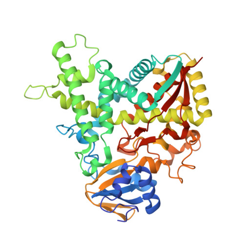
2VKU

4,4'-Dihydroxybenzophenone Mimics Sterol Substrate in the Binding Site of Sterol 14alpha-Demethylase (CYP51) in the X-ray Structure of the Complex


Experimental Data Snapshot

  • Method: X-RAY DIFFRACTION
  • Resolution: 1.95 Å
  • R-Value Free: 0.212 
  • R-Value Work: 0.177 
  • R-Value Observed: 0.177 

wwPDB Validation   3D Report Full Report


Ligand Structure Quality Assessment 


This is version 1.4 of the entry. See complete history


Literature

X-Ray Structure of 4,4'-Dihydroxybenzophenone Mimicking Sterol Substrate in the Active Site of Sterol 14Alpha-Demethylase (Cyp51)

Eddine, A.N.P Von Kries, J.Podust, M.V.Warrier, T.Kaufmann, S.H.E.Podust, L.M.

(2008) J Biol Chem 283: 15152

  • DOI: https://doi.org/10.1074/jbc.M801145200
  • Primary Citation of Related Structures:  
    2VKU

  • PubMed Abstract: 

    A universal step in the biosynthesis of membrane sterols and steroid hormones is the oxidative removal of the 14alpha-methyl group from sterol precursors by sterol 14alpha-demethylase (CYP51). This enzyme is a primary target in treatment of fungal infections in organisms ranging from humans to plants, and development of more potent and selective CYP51 inhibitors is an important biological objective. Our continuing interest in structural aspects of substrate and inhibitor recognition in CYP51 led us to determine (to a resolution of 1.95A) the structure of CYP51 from Mycobacterium tuberculosis (CYP51(Mt)) co-crystallized with 4,4'-dihydroxybenzophenone (DHBP), a small organic molecule previously identified among top type I binding hits in a library screened against CYP51(Mt). The newly determined CYP51(Mt)-DHBP structure is the most complete to date and is an improved template for three-dimensional modeling of CYP51 enzymes from fungal and prokaryotic pathogens. The structure demonstrates the induction of conformational fit of the flexible protein regions and the interactions of conserved Phe-89 essential for both fungal drug resistance and catalytic function, which were obscure in the previously characterized CYP51(Mt)-estriol complex. DHBP represents a benzophenone scaffold binding in the CYP51 active site via a type I mechanism, suggesting (i) a possible new class of CYP51 inhibitors targeting flexible regions, (ii) an alternative catalytic function for bacterial CYP51 enzymes, and (iii) a potential for hydroxybenzophenones, widely distributed in the environment, to interfere with sterol biosynthesis. Finally, we show the inhibition of M. tuberculosis growth by DHBP in a mouse macrophage model.


  • Organizational Affiliation

    Max-Planck-Institute for Infection Biology, Berlin, Germany.


Macromolecules
Find similar proteins by:  (by identity cutoff)  |  3D Structure
Entity ID: 1
MoleculeChains Sequence LengthOrganismDetailsImage
CYTOCHROME P450 51455Mycobacterium tuberculosis H37RvMutation(s): 2 
EC: 1.14.13.70
UniProt
Find proteins for P9WPP9 (Mycobacterium tuberculosis (strain ATCC 25618 / H37Rv))
Explore P9WPP9 
Go to UniProtKB:  P9WPP9
Entity Groups  
Sequence Clusters30% Identity50% Identity70% Identity90% Identity95% Identity100% Identity
UniProt GroupP9WPP9
Sequence Annotations
Expand
  • Reference Sequence
Small Molecules
Ligands 3 Unique
IDChains Name / Formula / InChI Key2D Diagram3D Interactions
HEM
Query on HEM

Download Ideal Coordinates CCD File 
F [auth A]PROTOPORPHYRIN IX CONTAINING FE
C34 H32 Fe N4 O4
KABFMIBPWCXCRK-RGGAHWMASA-L
DBE
Query on DBE

Download Ideal Coordinates CCD File 
B [auth A],
C [auth A],
D [auth A],
E [auth A]
bis(4-hydroxyphenyl)methanone
C13 H10 O3
RXNYJUSEXLAVNQ-UHFFFAOYSA-N
SO4
Query on SO4

Download Ideal Coordinates CCD File 
G [auth A]
H [auth A]
I [auth A]
J [auth A]
K [auth A]
G [auth A],
H [auth A],
I [auth A],
J [auth A],
K [auth A],
L [auth A]
SULFATE ION
O4 S
QAOWNCQODCNURD-UHFFFAOYSA-L
Binding Affinity Annotations 
IDSourceBinding Affinity
DBE BindingDB:  2VKU Kd: 2.80e+4 (nM) from 1 assay(s)
Binding MOAD:  2VKU Kd: 2.95e+4 (nM) from 1 assay(s)
Experimental Data & Validation

Experimental Data

  • Method: X-RAY DIFFRACTION
  • Resolution: 1.95 Å
  • R-Value Free: 0.212 
  • R-Value Work: 0.177 
  • R-Value Observed: 0.177 
  • Space Group: P 21 21 21
Unit Cell:
Length ( Å )Angle ( ˚ )
a = 77.734α = 90
b = 82.095β = 90
c = 85.859γ = 90
Software Package:
Software NamePurpose
CNSrefinement
HKL-2000data reduction
SCALEPACKdata scaling
MOLREPphasing

Structure Validation

View Full Validation Report


Ligand Structure Quality Assessment 


Entry History 

Deposition Data

Revision History  (Full details and data files)

  • Version 1.0: 2008-03-25
    Type: Initial release
  • Version 1.1: 2011-05-08
    Changes: Version format compliance
  • Version 1.2: 2011-07-13
    Changes: Version format compliance
  • Version 1.3: 2019-05-08
    Changes: Data collection, Experimental preparation, Other
  • Version 1.4: 2023-12-13
    Changes: Data collection, Database references, Derived calculations, Other, Refinement description