Conformational Changes and Reaction of Clostridial Glycosylating Toxins.
Ziegler, M.O.P., Jank, T., Aktories, K., Schulz, G.E.(2008) J Mol Biol 377: 1346
- PubMed: 18325534
- DOI: https://doi.org/10.1016/j.jmb.2007.12.065
- Primary Citation of Related Structures:
2VK9, 2VKD, 2VKH - PubMed Abstract:
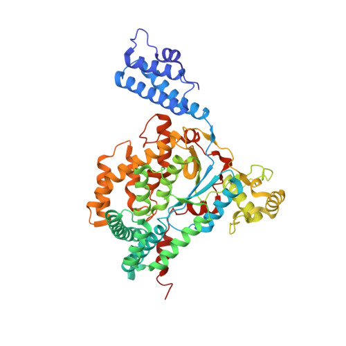
The crystal structures of the catalytic fragments of 'lethal toxin' from Clostridium sordellii and of 'alpha-toxin' from Clostridium novyi have been established. Almost half of the residues follow the chain fold of the glycosyl-transferase type A family of enzymes; the other half forms large alpha-helical protrusions that are likely to confer specificity for the respective targeted subgroup of Rho proteins in the cell. In the crystal, the active center of alpha-toxin contained no substrates and was disassembled, whereas that of lethal toxin, which was ligated with the donor substrate UDP-glucose and cofactor Mn2+, was catalytically competent. Surprisingly, the structure of lethal toxin with Ca2+ (instead of Mn2+) at the cofactor position showed a bound donor substrate with a disassembled active center, indicating that the strictly octahedral coordination sphere of Mn2+ is indispensable to the integrity of the enzyme. The homologous structures of alpha-toxin without substrate, distorted lethal toxin with Ca2+ plus donor, active lethal toxin with Mn2+ plus donor and the homologous Clostridium difficile toxin B with a hydrolyzed donor have been lined up to show the geometry of several reaction steps. Interestingly, the structural refinement of one of the three crystallographically independent molecules of Ca2+-ligated lethal toxin resulted in the glucosyl half-chair conformation expected for glycosyl-transferases that retain the anomeric configuration at the C1'' atom. A superposition of six acceptor substrates bound to homologous enzymes yielded the position of the nucleophilic acceptor atom with a deviation of <1 A. The resulting donor-acceptor geometry suggests that the reaction runs as a circular electron transfer in a six-membered ring, which involves the deprotonation of the nucleophile by the beta-phosphoryl group of the donor substrate UDP-glucose.
Organizational Affiliation:
Institut für Organische Chemie und Biochemie, Albert-Ludwigs-Universität, Albertstr. 21, D-79104 Freiburg im Breisgau, Germany.