2VJ8

Complex of human leukotriene A4 hydrolase with a hydroxamic acid inhibitor


Experimental Data Snapshot

  • Method: X-RAY DIFFRACTION
  • Resolution: 1.80 Å
  • R-Value Work: 0.195 
  • R-Value Observed: 0.195 

wwPDB Validation   3D Report Full Report


This is version 1.5 of the entry. See complete history


Literature

Crystal Structures of Leukotriene A4 Hydrolase in Complex with Captopril and Two Competitive Tight-Binding Inhibitors

Thunnissen, M.M.G.M.Andersson, B.Wong, C.-H.Samuelsson, B.Haeggstrom, J.Z.

(2002) FASEB J 16: 1648

  • DOI: https://doi.org/10.1096/fj.01-1017fje
  • Primary Citation of Related Structures:  
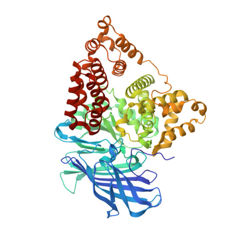
    2VJ8

  • PubMed Abstract: 

    Leukotriene (LT) A4 hydrolase/aminopeptidase is a bifunctional zinc enzyme that catalyzes the final step in the biosynthesis of LTB4, a potent chemoattractant and immune modulating lipid mediator. Here, we report a high-resolution crystal structure of LTA4 hydrolase in complex with captopril, a classical inhibitor of the zinc peptidase angiotensin-converting enzyme. Captopril makes few interactions with the protein, but its free thiol group is bound to the zinc, apparently accounting for most of its inhibitory action on LTA4 hydrolase. In addition, we have determined the structures of LTA4 hydrolase in complex with two selective tight-binding inhibitors, a thioamine and a hydroxamic acid. Their common benzyloxyphenyl tail, designed to mimic the carbon backbone of LTA4, binds into a narrow hydrophobic cavity in the protein. The free hydroxyl group of the hydroxamic acid makes a suboptimal, monodentate complex with the zinc, and strategies for improved inhibitor design can be deduced from the structure. Taken together, the three crystal structures provide the molecular basis for the divergent pharmacological profiles of LTA4 hydrolase inhibitors. Moreover, they help define the binding pocket for the fatty acid-derived epoxide LTA4 as well as the subsites for a tripeptide substrate, which in turn have important implications for the molecular mechanisms of enzyme catalyses.


  • Organizational Affiliation

    Department of Biochemistry, University of Stockholm, Arrhenius Laboratories A4, S-106 91 Stockholm, Sweden.


Macromolecules
Find similar proteins by:  (by identity cutoff)  |  3D Structure
Entity ID: 1
MoleculeChains Sequence LengthOrganismDetailsImage
LTA4H PROTEIN611Homo sapiensMutation(s): 0 
EC: 3.3.2.6
UniProt & NIH Common Fund Data Resources
Find proteins for P09960 (Homo sapiens)
Explore P09960 
Go to UniProtKB:  P09960
PHAROS:  P09960
GTEx:  ENSG00000111144 
Entity Groups  
Sequence Clusters30% Identity50% Identity70% Identity90% Identity95% Identity100% Identity
UniProt GroupP09960
Sequence Annotations
Expand
  • Reference Sequence
Small Molecules
Ligands 5 Unique
IDChains Name / Formula / InChI Key2D Diagram3D Interactions
HA2
Query on HA2

Download Ideal Coordinates CCD File 
G [auth A]6-[{(2S)-2-AMINO-3-[4-(BENZYLOXY)PHENYL]PROPYL}(HYDROXY)AMINO]-6-OXOHEXANOIC ACID)
C22 H28 N2 O5
YOYHDDAKAGRLRC-IBGZPJMESA-N
YB
Query on YB

Download Ideal Coordinates CCD File 
C [auth A],
D [auth A],
H [auth A]
YTTERBIUM (III) ION
Yb
AWSFICBXMUKWSK-UHFFFAOYSA-N
IMD
Query on IMD

Download Ideal Coordinates CCD File 
E [auth A]IMIDAZOLE
C3 H5 N2
RAXXELZNTBOGNW-UHFFFAOYSA-O
ZN
Query on ZN

Download Ideal Coordinates CCD File 
B [auth A]ZINC ION
Zn
PTFCDOFLOPIGGS-UHFFFAOYSA-N
ACT
Query on ACT

Download Ideal Coordinates CCD File 
F [auth A]ACETATE ION
C2 H3 O2
QTBSBXVTEAMEQO-UHFFFAOYSA-M
Binding Affinity Annotations 
IDSourceBinding Affinity
HA2 Binding MOAD:  2VJ8 Ki: 2 (nM) from 1 assay(s)
PDBBind:  2VJ8 Ki: 2 (nM) from 1 assay(s)
Experimental Data & Validation

Experimental Data

  • Method: X-RAY DIFFRACTION
  • Resolution: 1.80 Å
  • R-Value Work: 0.195 
  • R-Value Observed: 0.195 
  • Space Group: P 21 21 2
Unit Cell:
Length ( Å )Angle ( ˚ )
a = 67.77α = 90
b = 132.47β = 90
c = 83.7γ = 90
Software Package:
Software NamePurpose
TNTrefinement
MOSFLMdata reduction
SCALEPACKdata scaling

Structure Validation

View Full Validation Report


Entry History 

Deposition Data

Revision History  (Full details and data files)

  • Version 1.0: 2008-01-15
    Type: Initial release
  • Version 1.1: 2011-05-08
    Changes: Version format compliance
  • Version 1.2: 2011-07-13
    Changes: Version format compliance
  • Version 1.3: 2018-01-17
    Changes: Data collection
  • Version 1.4: 2019-02-27
    Changes: Data collection, Experimental preparation
  • Version 1.5: 2023-12-13
    Changes: Data collection, Database references, Derived calculations, Refinement description