


Crystal Structure of Human Short-Chain Acyl Coa Dehydrogenase
Pike, A.C.W., Pantic, N., Parizotto, E., Gileadi, O., Ugochukwu, E., von Delft, F., Weigelt, J., Arrowsmith, C.H., Edwards, A., Oppermann, U.To be published.
Experimental Data Snapshot
Entity ID: 1 | |||||
|---|---|---|---|---|---|
| Molecule | Chains | Sequence Length | Organism | Details | Image |
| SHORT-CHAIN SPECIFIC ACYL-COA DEHYDROGENASE, | 391 | Homo sapiens | Mutation(s): 0 EC: 1.3.99.2 | 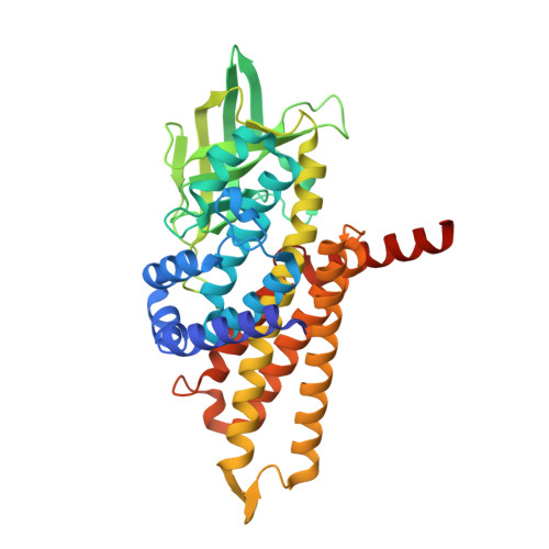 | |
UniProt & NIH Common Fund Data Resources | |||||
Find proteins for P16219 (Homo sapiens) Explore P16219 Go to UniProtKB: P16219 | |||||
PHAROS: P16219 GTEx: ENSG00000122971 | |||||
Entity Groups | |||||
| Sequence Clusters | 30% Identity50% Identity70% Identity90% Identity95% Identity100% Identity | ||||
| UniProt Group | P16219 | ||||
Sequence AnnotationsExpand | |||||
| |||||
| Ligands 3 Unique | |||||
|---|---|---|---|---|---|
| ID | Chains | Name / Formula / InChI Key | 2D Diagram | 3D Interactions | |
| COS Query on COS | DA [auth G], L [auth B], O [auth C], Q [auth D], Y [auth F] | COENZYME A PERSULFIDE C21 H36 N7 O16 P3 S2 REVPHPVBPSIEKM-IBOSZNHHSA-N |  | ||
| FAD Query on FAD | CA [auth G] EA [auth H] I [auth A] K [auth B] N [auth C] | FLAVIN-ADENINE DINUCLEOTIDE C27 H33 N9 O15 P2 VWWQXMAJTJZDQX-UYBVJOGSSA-N |  | ||
| EDO Query on EDO | AA [auth F] BA [auth F] FA [auth H] GA [auth H] J [auth A] | 1,2-ETHANEDIOL C2 H6 O2 LYCAIKOWRPUZTN-UHFFFAOYSA-N |  | ||
| Length ( Å ) | Angle ( ˚ ) |
|---|---|
| a = 85.714 | α = 90 |
| b = 157.62 | β = 90 |
| c = 260.843 | γ = 90 |
| Software Name | Purpose |
|---|---|
| REFMAC | refinement |
| XDS | data reduction |
| XSCALE | data scaling |
| PHASER | phasing |
RCSB PDB (citation) is hosted by
RCSB PDB is a member of the

