2VHL

The Three-dimensional structure of the N-Acetylglucosamine-6- phosphate deacetylase from Bacillus subtilis


Experimental Data Snapshot

  • Method: X-RAY DIFFRACTION
  • Resolution: 2.05 Å
  • R-Value Free: 0.260 
  • R-Value Work: 0.205 
  • R-Value Observed: 0.208 

wwPDB Validation   3D Report Full Report


Ligand Structure Quality Assessment 


This is version 1.5 of the entry. See complete history


Literature

The Three-Dimensional Structure of the N-Acetylglucosamine-6-Phosphate Deacetylase from Bacillus Subtilis

Vincent, F.Yates, D.Garman, E.Davies, G.J.

(2004) J Biol Chem 279: 2809

  • DOI: https://doi.org/10.1074/jbc.M310165200
  • Primary Citation of Related Structures:  
    2VHL

  • PubMed Abstract: 

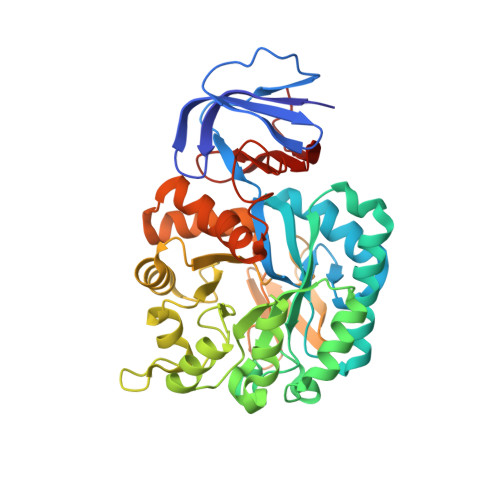
    The enzyme N-acetylglucosamine-6-phosphate deacetylase, NagA, catalyzes the hydrolysis of the N-acetyl group of GlcNAc-6-P to yield glucosamine 6-phosphate and acetate, the first committed step in the biosynthetic pathway to amino-sugar-nucleotides. It is classified into carbohydrate esterase family CE-9 (see afmb.cnrs-mrs.fr/CAZY/). Here we report the cloning, expression, and three-dimensional structure (Protein Data Bank code 1un7) determination by x-ray crystallography of the Bacillus subtilis NagA at a resolution of 2.0 A. The structure presents two domains, a (beta/alpha)(8) barrel enclosing the active center and a small beta barrel domain. The structure is dimeric, and the substrate phosphate coordination at the active center is provided by an Arg/His pair contributed from the second molecule of the dimer. Both the overall structure and the active center bear a striking similarity to the urease superfamily with two metals involved in substrate binding and catalysis. PIXE (Proton-Induced x-ray Emission) data show that iron is the predominant metal in the purified protein. We propose a catalytic mechanism involving proton donation to the leaving group by aspartate, nucleophilic attack by an Fe-bridged hydroxide, and stabilization of the carbonyl oxygen by one of the two Fe atoms of the pair. We believe that this is the first sugar deacetylase to utilize this fold and catalytic mechanism.


  • Organizational Affiliation

    Department of Chemistry, The University of York, Heslington, York, YO10 5YW, United Kingdom.


Macromolecules
Find similar proteins by:  (by identity cutoff)  |  3D Structure
Entity ID: 1
MoleculeChains Sequence LengthOrganismDetailsImage
N-ACETYLGLUCOSAMINE-6-PHOSPHATE DEACETYLASE
A, B
396Bacillus subtilisMutation(s): 0 
EC: 3.5.1.25
UniProt
Find proteins for O34450 (Bacillus subtilis (strain 168))
Explore O34450 
Go to UniProtKB:  O34450
Entity Groups  
Sequence Clusters30% Identity50% Identity70% Identity90% Identity95% Identity100% Identity
UniProt GroupO34450
Sequence Annotations
Expand
  • Reference Sequence
Small Molecules
Ligands 3 Unique
IDChains Name / Formula / InChI Key2D Diagram3D Interactions
GLP
Query on GLP

Download Ideal Coordinates CCD File 
C [auth A],
G [auth B]
2-amino-2-deoxy-6-O-phosphono-alpha-D-glucopyranose
C6 H14 N O8 P
XHMJOUIAFHJHBW-UKFBFLRUSA-N
PGE
Query on PGE

Download Ideal Coordinates CCD File 
D [auth A],
H [auth B]
TRIETHYLENE GLYCOL
C6 H14 O4
ZIBGPFATKBEMQZ-UHFFFAOYSA-N
FE
Query on FE

Download Ideal Coordinates CCD File 
E [auth A],
F [auth A],
I [auth B],
J [auth B]
FE (III) ION
Fe
VTLYFUHAOXGGBS-UHFFFAOYSA-N
Experimental Data & Validation

Experimental Data

  • Method: X-RAY DIFFRACTION
  • Resolution: 2.05 Å
  • R-Value Free: 0.260 
  • R-Value Work: 0.205 
  • R-Value Observed: 0.208 
  • Space Group: P 21 21 2
Unit Cell:
Length ( Å )Angle ( ˚ )
a = 51.68α = 90
b = 107.74β = 90
c = 188.25γ = 90
Software Package:
Software NamePurpose
REFMACrefinement
AMoREphasing

Structure Validation

View Full Validation Report


Ligand Structure Quality Assessment 


Entry History 

Deposition Data

Revision History  (Full details and data files)

  • Version 1.0: 2007-12-04
    Type: Initial release
  • Version 1.1: 2011-05-08
    Changes: Version format compliance
  • Version 1.2: 2011-07-13
    Changes: Version format compliance
  • Version 1.3: 2020-07-29
    Type: Remediation
    Reason: Carbohydrate remediation
    Changes: Data collection, Derived calculations, Other, Structure summary
  • Version 1.4: 2021-03-24
    Changes: Derived calculations, Structure summary
  • Version 1.5: 2023-12-13
    Changes: Data collection, Database references, Refinement description